Copyright
©The Author(s) 2015.
World J Gastroenterol. Sep 7, 2015; 21(33): 9774-9784
Published online Sep 7, 2015. doi: 10.3748/wjg.v21.i33.9774
Published online Sep 7, 2015. doi: 10.3748/wjg.v21.i33.9774
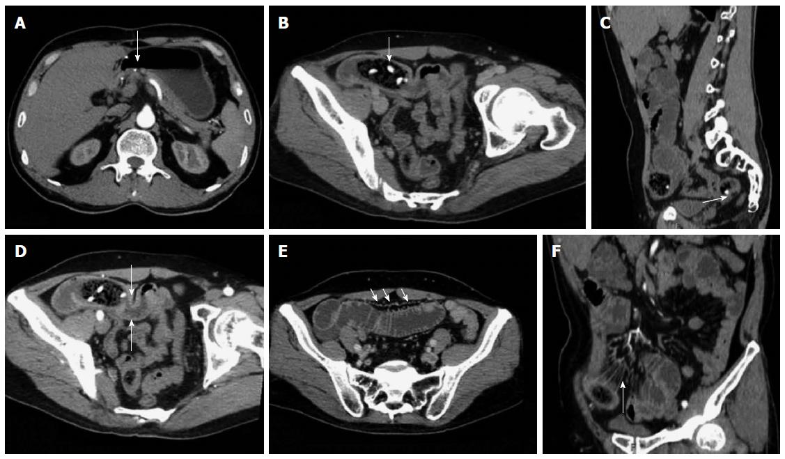
Figure 3 Representative case of a 58-year-old male with hawthorn small bowel obstruction.
A: Contrast-enhanced examination result displaying the gastroduodenal anastomotic stoma of the artery; B: Oblique MPR revealing an oval bezoar in the distal end of the jejunum with shadows of high-density seeds (arrow); C: Sagittal reconstruction showing co-existing bezoar inside the ileum (arrow); D: Arterial phase image exhibiting the thickened and strengthened intestinal wall in the distal end of the obstruction site (arrow); E: Portal venous phase image showing a significantly enlarged vascular shadow and blurred mesentery of the proximal end of the dilated SBO intestine (arrow); F: CTA image revealing the thickening of the mesenteric blood vessel and peripheral exudation in the obstruction site (arrow).
- Citation: Wang PY, Wang X, Zhang L, Li HF, Chen L, Wang X, Wang B. Bezoar-induced small bowel obstruction: Clinical characteristics and diagnostic value of multi-slice spiral computed tomography. World J Gastroenterol 2015; 21(33): 9774-9784
- URL: https://www.wjgnet.com/1007-9327/full/v21/i33/9774.htm
- DOI: https://dx.doi.org/10.3748/wjg.v21.i33.9774


