Copyright
©The Author(s) 2015.
World J Gastroenterol. Aug 28, 2015; 21(32): 9494-9502
Published online Aug 28, 2015. doi: 10.3748/wjg.v21.i32.9494
Published online Aug 28, 2015. doi: 10.3748/wjg.v21.i32.9494
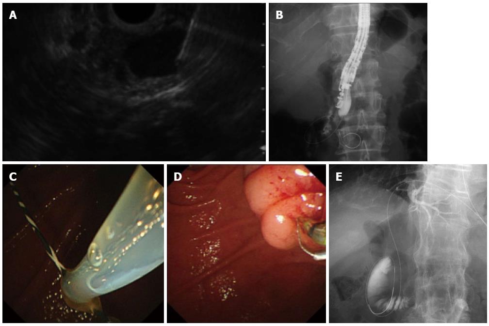
Figure 4 Endoscopic ultrasonography-guided rendezvous procedure.
A: Endoscopic ultrasonography showing that extrahepatic bile duct was punctured by the needle; B: Radiography showing the guidewire was passed through the needle into the duodenum across the papilla antegradely. The endoscope was in the short position; C: Following echoendoscope withdrawal, the duodenoscopic view showed that a guidewire passing through the papilla was grasped by the snare; D: After the guidewire was pulled out through the accessary channel of the duodenoscope, a catheter was inserted into the bile duct over the existing guidewire; E: Transhepatic approach. Abdominal radiography shows that a guidewire was placed through the papilla into the duodenum.
- Citation: Kawakubo K, Kawakami H, Kuwatani M, Haba S, Kawahata S, Abe Y, Kubota Y, Kubo K, Isayama H, Sakamoto N. Recent advances in endoscopic ultrasonography-guided biliary interventions. World J Gastroenterol 2015; 21(32): 9494-9502
- URL: https://www.wjgnet.com/1007-9327/full/v21/i32/9494.htm
- DOI: https://dx.doi.org/10.3748/wjg.v21.i32.9494


