Copyright
©The Author(s) 2015.
World J Gastroenterol. Aug 28, 2015; 21(32): 9494-9502
Published online Aug 28, 2015. doi: 10.3748/wjg.v21.i32.9494
Published online Aug 28, 2015. doi: 10.3748/wjg.v21.i32.9494
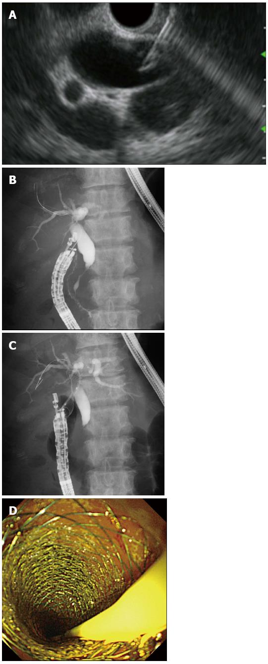
Figure 1 Endoscopic ultrasonography-guided choledochoduodenostomy.
A: Endoscopic ultrasonography showing that extrahepatic bile duct was punctured by the needle; B: Guidewire was advanced through the needle into the right liver lobe; C: A self-expandable metallic stent (SEMS) was placed between the bile duct and the duodenum; D: Endoscopic view showing that the distal end of a SEMS was located at the duodenum.
- Citation: Kawakubo K, Kawakami H, Kuwatani M, Haba S, Kawahata S, Abe Y, Kubota Y, Kubo K, Isayama H, Sakamoto N. Recent advances in endoscopic ultrasonography-guided biliary interventions. World J Gastroenterol 2015; 21(32): 9494-9502
- URL: https://www.wjgnet.com/1007-9327/full/v21/i32/9494.htm
- DOI: https://dx.doi.org/10.3748/wjg.v21.i32.9494


