Copyright
©The Author(s) 2015.
World J Gastroenterol. Aug 21, 2015; 21(31): 9437-9441
Published online Aug 21, 2015. doi: 10.3748/wjg.v21.i31.9437
Published online Aug 21, 2015. doi: 10.3748/wjg.v21.i31.9437
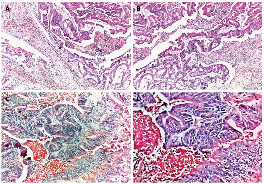
Figure 3 Histological findings of surgical specimen demonstrating moderately differentiated adenocarcinoma, infiltrating the wall thickness (A) (HE, magnification × 5), with areas of cribriform appearance due to fusion of glands and areas of necrosis (B) (HE, magnification × 10); A higher magnification, demonstrating dysplastic aspect of epithelium, loss of polarity and cell dysplasia (C) (HE, magnification × 100) and (D) (HE, magnification × 200).
HE: Hematoxylin and eosin.
- Citation: Paquissi FC, Lima AHFBP, Lopes MFDNV, Diaz FV. Adenocarcinoma of the third and fourth portions of the duodenum: The capsule endoscopy value. World J Gastroenterol 2015; 21(31): 9437-9441
- URL: https://www.wjgnet.com/1007-9327/full/v21/i31/9437.htm
- DOI: https://dx.doi.org/10.3748/wjg.v21.i31.9437


