Copyright
©The Author(s) 2015.
World J Gastroenterol. Aug 21, 2015; 21(31): 9358-9366
Published online Aug 21, 2015. doi: 10.3748/wjg.v21.i31.9358
Published online Aug 21, 2015. doi: 10.3748/wjg.v21.i31.9358
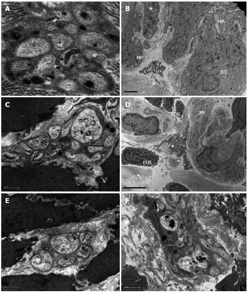
Figure 1 Transmission electron microscopy of cathartic colons.
In the colons of control animals, normal A: Nerve fibers (Nf; magnification × 40000); and B: Interstitial cells of Cajal (ICC; magnification × 2500) can be seen. In contrast, cathartic colons in model animals showed C: Vacuole formation in ‘mit’ and sparse neurofilament (magnification × 11500); D: Eosinophil (EOS) infiltration (magnification × 1750); E (magnification × 5900) and F (magnification × 5900) each for mosapride and FAI showing recovery of damage.
- Citation: Wang SY, Liu YP, Fan YH, Zhang L, Cai LJ, Lv B. Mechanism of aqueous fructus aurantii immaturus extracts in neuroplexus of cathartic colons. World J Gastroenterol 2015; 21(31): 9358-9366
- URL: https://www.wjgnet.com/1007-9327/full/v21/i31/9358.htm
- DOI: https://dx.doi.org/10.3748/wjg.v21.i31.9358


