Copyright
©The Author(s) 2015.
World J Gastroenterol. Aug 14, 2015; 21(30): 9217-9222
Published online Aug 14, 2015. doi: 10.3748/wjg.v21.i30.9217
Published online Aug 14, 2015. doi: 10.3748/wjg.v21.i30.9217
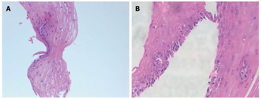
Figure 5 Microscopy.
Esophageal mucosa with dyskeratotic squamous cells, apoptosis of individual squamous cells, acanthosis and spongiosis. Hematoxylin-eosin staining, magnification × 100 (A), × 200 (B), respectively.
-
Citation: Trabulo D, Ferreira S, Lage P, Rego RL, Teixeira G, Pereira AD. Esophageal stenosis with sloughing esophagitis: A curious manifestation of graft-
vs -host disease. World J Gastroenterol 2015; 21(30): 9217-9222 - URL: https://www.wjgnet.com/1007-9327/full/v21/i30/9217.htm
- DOI: https://dx.doi.org/10.3748/wjg.v21.i30.9217


