Copyright
©The Author(s) 2015.
World J Gastroenterol. Jan 21, 2015; 21(3): 988-996
Published online Jan 21, 2015. doi: 10.3748/wjg.v21.i3.988
Published online Jan 21, 2015. doi: 10.3748/wjg.v21.i3.988
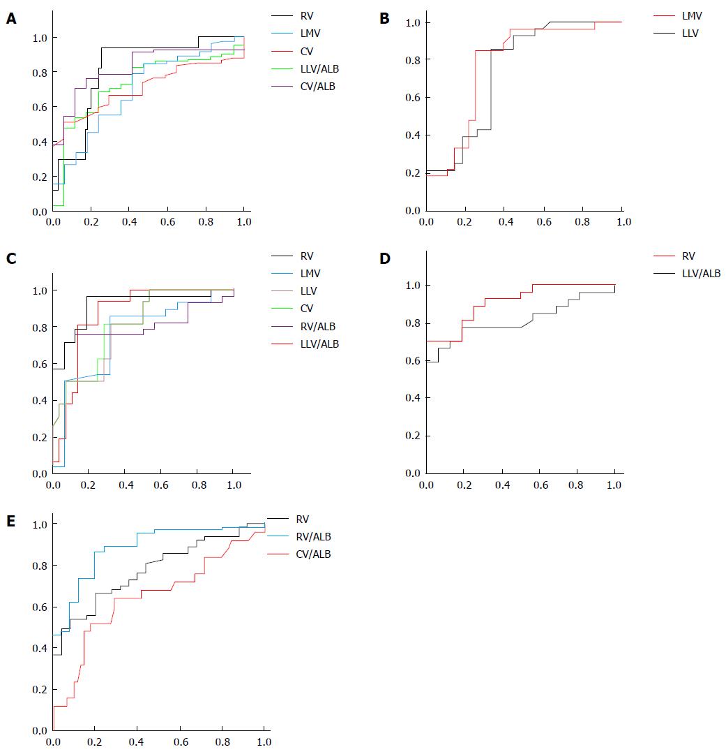
Figure 2 Receiver operating characteristic curves of liver lobe volume parameters to identify the presence, and Child-Pugh class, of liver cirrhosis in patients with hepatitis B, and the presence of esophageal varices.
The figures show that the ratio of caudate lobe volume to albumin (CV/ALB), left lateral liver lobe volume (LLV), right liver lobe volume (RV), the ratio of LLV to albumin (LLV/ALB) and the ratio of RV to albumin (RV/ALB) could be recommended as an indicator for distinguishing cirrhotic patients from healthy participants (A), Child-Pugh class A from B (B), class A from C (C), class B from C (D), and cirrhotic patients with esophageal varices from those without esophageal varices (E), respectively.
- Citation: Li H, Chen TW, Li ZL, Zhang XM, Li CJ, Chen XL, Chen GW, Hu JN, Ye YQ. Albumin and magnetic resonance imaging-liver volume to identify hepatitis B-related cirrhosis and esophageal varices. World J Gastroenterol 2015; 21(3): 988-996
- URL: https://www.wjgnet.com/1007-9327/full/v21/i3/988.htm
- DOI: https://dx.doi.org/10.3748/wjg.v21.i3.988


