Copyright
©The Author(s) 2015.
World J Gastroenterol. Jan 21, 2015; 21(3): 888-896
Published online Jan 21, 2015. doi: 10.3748/wjg.v21.i3.888
Published online Jan 21, 2015. doi: 10.3748/wjg.v21.i3.888
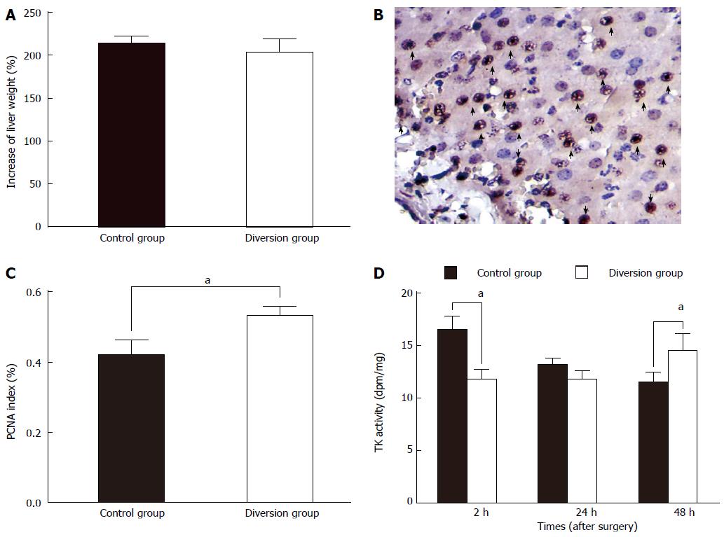
Figure 6 This indicated that the hyperperfusion could be relieved in a short time, which can be attributed to rapid regeneration of the liver remnant after massive hepatectomy.
A: Increase rate of the liver remnant in both groups at 48 h PH (post hepatectomy). There was no significant difference between the control and diversion groups (P = NS); B: Proliferation cell nuclear antigen (PCNA) staining in the liver remnant (positive cells identified by dark-brown-stained nuclei, indicated by arrows; magnification × 400); C: Microphotometric evaluation of PCNA index in PCNA-stained tissue at 48 h PH. Values are expressed as means ± SD for both groups; D: The change in thymidine kinase (TK) activity in both groups. TK activity in the control group was significantly elevated compared with the diversion group at 2 h PH (P = 0.04), but there was no significant difference between the groups at 24 h PH (P = NS). However, TK activity in the control group was significantly reduced compared to that in the diversion group at 48 h PH. aP < 0.05, diversion group vs control group. NS: Not significant.
- Citation: Wang DD, Xu Y, Zhu ZM, Tan XL, Tu YL, Han MM, Tan JW. Should temporary extracorporeal continuous portal diversion replace meso/porta-caval shunts in “small-for-size” syndrome in porcine hepatectomy? World J Gastroenterol 2015; 21(3): 888-896
- URL: https://www.wjgnet.com/1007-9327/full/v21/i3/888.htm
- DOI: https://dx.doi.org/10.3748/wjg.v21.i3.888


