Copyright
©The Author(s) 2015.
World J Gastroenterol. Jan 21, 2015; 21(3): 862-867
Published online Jan 21, 2015. doi: 10.3748/wjg.v21.i3.862
Published online Jan 21, 2015. doi: 10.3748/wjg.v21.i3.862
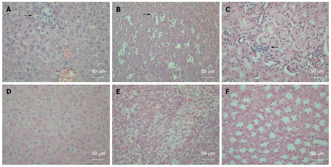
Figure 3 Histopathologic changes in the hepatitis E virus-inoculated Mongolian gerbils.
A: A representative liver sample showing slight histiocytic hepatitis and focal accumulation of inflammatory cells surrounding hepatocytes [21 d post-inoculation (DPI)]; B: A representative spleen sample showing ruptured and enlarged splenocytes with multiple vacuolar degeneration (21 DPI); C: A representative kidney showing disarranged kidney cells with increased infiltrating lymphocytes and macrophages (35 DPI); D: A representative negative control liver; E: A representative negative control spleen; F: A representative Negative control kidney. All tissues were stained with hematoxylin and eosin. Original magnifications × 400.
- Citation: Hong Y, He ZJ, Tao W, Fu T, Wang YK, Chen Y. Experimental infection of Z:ZCLA Mongolian gerbils with human hepatitis E virus. World J Gastroenterol 2015; 21(3): 862-867
- URL: https://www.wjgnet.com/1007-9327/full/v21/i3/862.htm
- DOI: https://dx.doi.org/10.3748/wjg.v21.i3.862


