Copyright
©The Author(s) 2015.
World J Gastroenterol. Jan 21, 2015; 21(3): 759-785
Published online Jan 21, 2015. doi: 10.3748/wjg.v21.i3.759
Published online Jan 21, 2015. doi: 10.3748/wjg.v21.i3.759
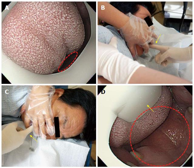
Figure 8 Intubation of scope from oral cavity into pharynx.
A: Tongue blocking the oral cavity. As the oral cavity has a very narrow space (dotted circle) in this case, it is difficult to insert the scope (B, C). The examiner pushes aside the examinee’s tongue with his or her index finger (arrow mark) to secure more space; D: Tongue placed under the tongue restrainer (arrow mark). After securing more space, the insertion of the scope is easier.
- Citation: Lee SH, Park YK, Cho SM, Kang JK, Lee DJ. Technical skills and training of upper gastrointestinal endoscopy for new beginners. World J Gastroenterol 2015; 21(3): 759-785
- URL: https://www.wjgnet.com/1007-9327/full/v21/i3/759.htm
- DOI: https://dx.doi.org/10.3748/wjg.v21.i3.759


