Copyright
©The Author(s) 2015.
World J Gastroenterol. Jan 21, 2015; 21(3): 1036-1039
Published online Jan 21, 2015. doi: 10.3748/wjg.v21.i3.1036
Published online Jan 21, 2015. doi: 10.3748/wjg.v21.i3.1036
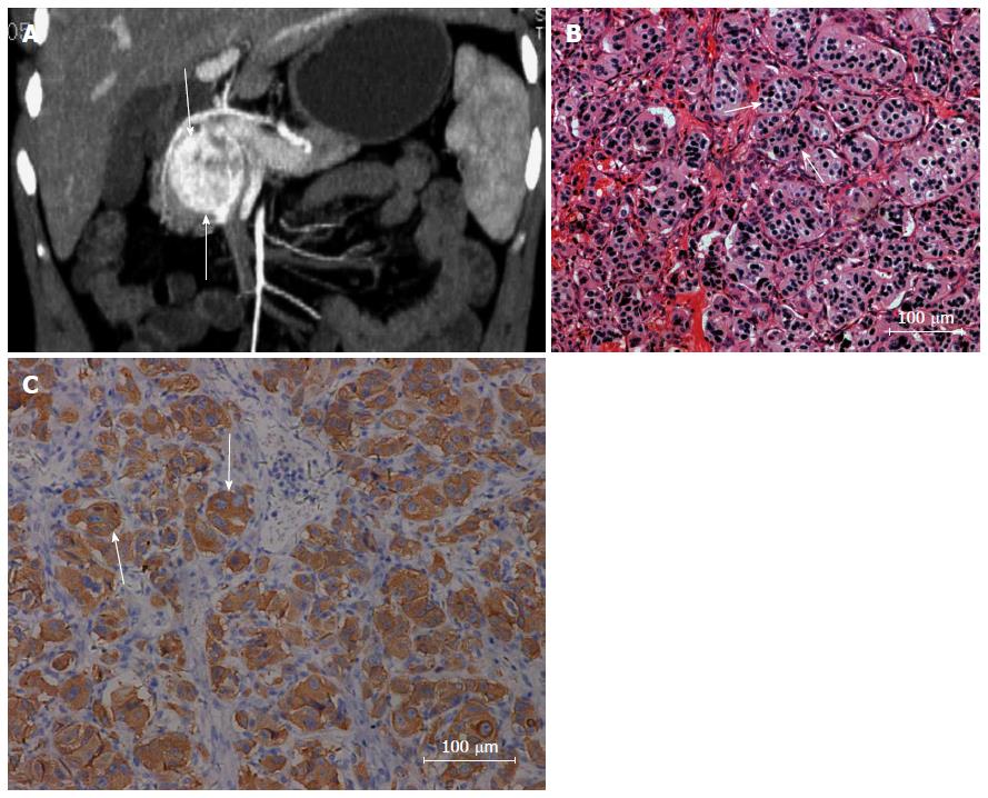
Figure 2 Forty-one-year-old woman was found to have an upper abdominal mass on ultrasound examination 6 d earlier.
A: On coronal reconstruction imaging, a well-demarcated mass (arrows) was found with marked enhancement in the arterial phase after contrast enhancement; B: Under microscopic examination, a few cells had large nuclei with hyperchromatism and pink cytoplasm, arranged in organ-like or nest-like structures (arrows) (hematoxylin-eosin; original magnification, 200 ×); C: Immunohistochemistry showed positive staining for synaptophysin (200 ×).
- Citation: Meng L, Wang J, Fang SH. Primary pancreatic paraganglioma: A report of two cases and literature review. World J Gastroenterol 2015; 21(3): 1036-1039
- URL: https://www.wjgnet.com/1007-9327/full/v21/i3/1036.htm
- DOI: https://dx.doi.org/10.3748/wjg.v21.i3.1036


