Copyright
©The Author(s) 2015.
World J Gastroenterol. Jan 21, 2015; 21(3): 1036-1039
Published online Jan 21, 2015. doi: 10.3748/wjg.v21.i3.1036
Published online Jan 21, 2015. doi: 10.3748/wjg.v21.i3.1036
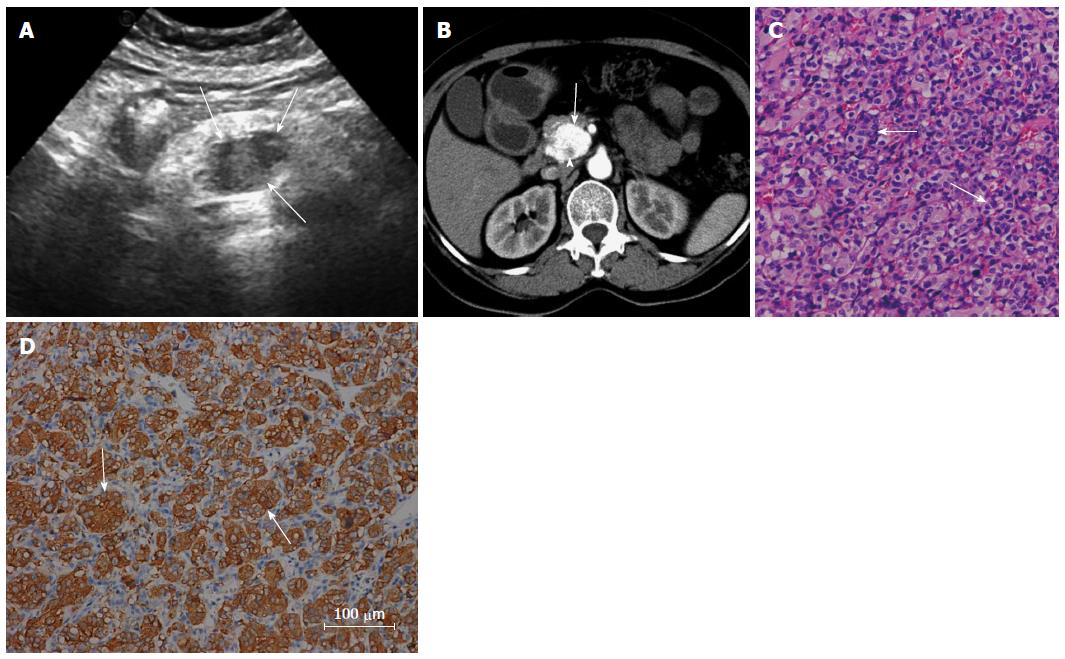
Figure 1 Fifty-four-year-old woman felt left upper abdominal pain for 20 d.
A: Ultrasonography revealed an ill-defined hypoechoic mass in the pancreatic head (arrows); B: On contrast-enhanced computed tomography, the mass (arrow) in the pancreatic head was found with heterogeneous marked enhancement in the arterial phase, while non-enhancing patchy areas were seen in the tumor (arrowhead); C: Microscopically, the chief cells had round or ovoid nuclei (arrows), arranged in nest-like structures with dilated capillary intervals (hematoxylin-eosin; original magnification, 200 ×); D: Immunohistochemical study revealed positive staining for synaptophysin (200 ×).
- Citation: Meng L, Wang J, Fang SH. Primary pancreatic paraganglioma: A report of two cases and literature review. World J Gastroenterol 2015; 21(3): 1036-1039
- URL: https://www.wjgnet.com/1007-9327/full/v21/i3/1036.htm
- DOI: https://dx.doi.org/10.3748/wjg.v21.i3.1036


