Copyright
©The Author(s) 2015.
World J Gastroenterol. Aug 7, 2015; 21(29): 8974-8980
Published online Aug 7, 2015. doi: 10.3748/wjg.v21.i29.8974
Published online Aug 7, 2015. doi: 10.3748/wjg.v21.i29.8974
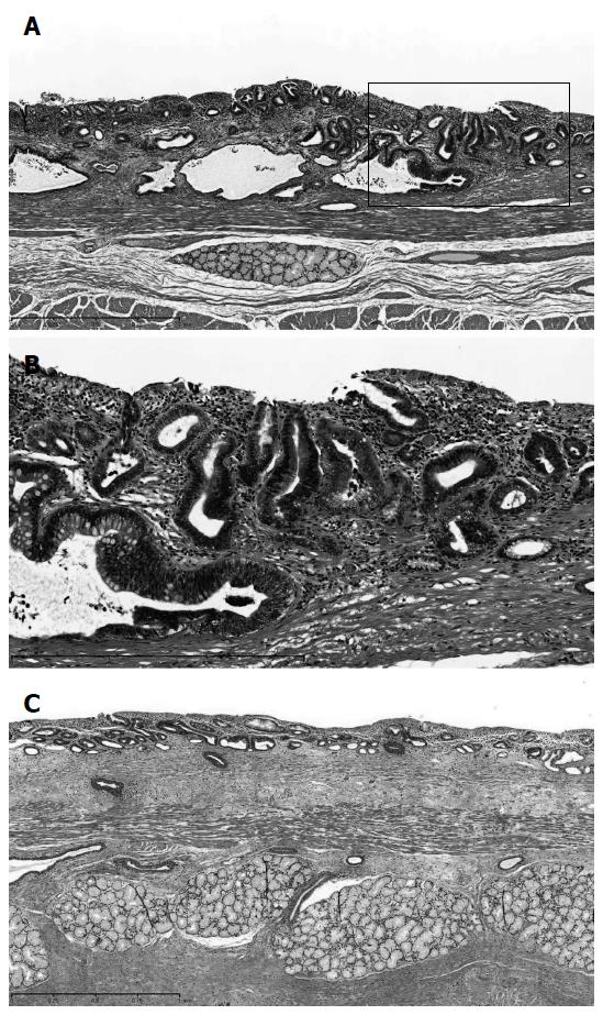
Figure 5 Histopathological examination.
A: A histopathological examination revealed superficial adenocarcinoma (pT1a-SMM). The cancer lesion was surrounded by mucosa, including a columnar epithelium that continued to the stomach; B: Part of the small frame in (A) was magnified and shown; C: Duplication of the muscularis mucosae was identified in long-segment Barrett's esophagus. Hematoxylin-eosin staining; magnification, × 100 (A), × 150 (B) or × 50 (C).
- Citation: Shiozaki A, Fujiwara H, Konishi H, Kinoshita O, Kosuga T, Morimura R, Murayama Y, Komatsu S, Kuriu Y, Ikoma H, Nakanishi M, Ichikawa D, Okamoto K, Sakakura C, Otsuji E. Laparoscopic transhiatal approach for resection of an adenocarcinoma in long-segment Barrett’s esophagus. World J Gastroenterol 2015; 21(29): 8974-8980
- URL: https://www.wjgnet.com/1007-9327/full/v21/i29/8974.htm
- DOI: https://dx.doi.org/10.3748/wjg.v21.i29.8974


