Copyright
©The Author(s) 2015.
World J Gastroenterol. Aug 7, 2015; 21(29): 8974-8980
Published online Aug 7, 2015. doi: 10.3748/wjg.v21.i29.8974
Published online Aug 7, 2015. doi: 10.3748/wjg.v21.i29.8974
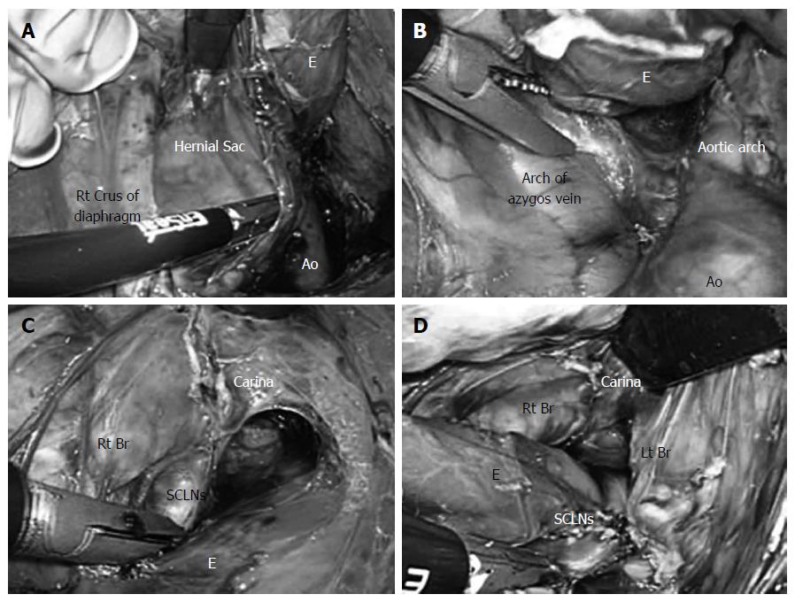
Figure 3 Thoracic esophagus was completely detached from the surrounding tissue.
A: A hernial sac was identified on the cranial side of the right crus of the diaphragm; B: Dissection of the posterior and right sides of the esophagus was performed to the level of the arch of the azygos vein; C: While lifting the right mediastinal pleura like a membrane, an incision was made and extended to the right pulmonary hilum, and the lymph nodes were resected from the right main bronchus and carina; D: Intraoperative view after dissection of the subcarinal lymph nodes. Ao: Thoracic aorta; E: Esophagus; Lt: Left; Rt: Right; Br: Bronchus; SCLNs: Subcarinal lymph nodes.
- Citation: Shiozaki A, Fujiwara H, Konishi H, Kinoshita O, Kosuga T, Morimura R, Murayama Y, Komatsu S, Kuriu Y, Ikoma H, Nakanishi M, Ichikawa D, Okamoto K, Sakakura C, Otsuji E. Laparoscopic transhiatal approach for resection of an adenocarcinoma in long-segment Barrett’s esophagus. World J Gastroenterol 2015; 21(29): 8974-8980
- URL: https://www.wjgnet.com/1007-9327/full/v21/i29/8974.htm
- DOI: https://dx.doi.org/10.3748/wjg.v21.i29.8974


