Copyright
©The Author(s) 2015.
World J Gastroenterol. Aug 7, 2015; 21(29): 8974-8980
Published online Aug 7, 2015. doi: 10.3748/wjg.v21.i29.8974
Published online Aug 7, 2015. doi: 10.3748/wjg.v21.i29.8974
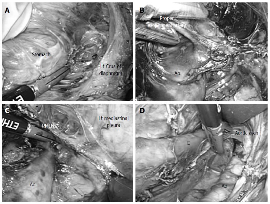
Figure 2 Surgical technique.
A: The esophageal hiatus was divided, and carbon dioxide was introduced into the mediastinum; B: Dissection of the anterior plane of the thoracic aorta was extended to the cranial side, and the root of the proper esophageal artery was confirmed under a magnified videoscopic view; C: While lifting the posterior mediastinal lymph nodes like a membrane, they were cut along the border of the left mediastinal pleura; D: This incision was extended to the left pulmonary hilum and aortic arch. Ao: Thoracic aorta; E: Esophagus; Lt: Left; PMLNs: Posterior mediastinal lymph nodes.
- Citation: Shiozaki A, Fujiwara H, Konishi H, Kinoshita O, Kosuga T, Morimura R, Murayama Y, Komatsu S, Kuriu Y, Ikoma H, Nakanishi M, Ichikawa D, Okamoto K, Sakakura C, Otsuji E. Laparoscopic transhiatal approach for resection of an adenocarcinoma in long-segment Barrett’s esophagus. World J Gastroenterol 2015; 21(29): 8974-8980
- URL: https://www.wjgnet.com/1007-9327/full/v21/i29/8974.htm
- DOI: https://dx.doi.org/10.3748/wjg.v21.i29.8974


