Copyright
©The Author(s) 2015.
World J Gastroenterol. Aug 7, 2015; 21(29): 8974-8980
Published online Aug 7, 2015. doi: 10.3748/wjg.v21.i29.8974
Published online Aug 7, 2015. doi: 10.3748/wjg.v21.i29.8974
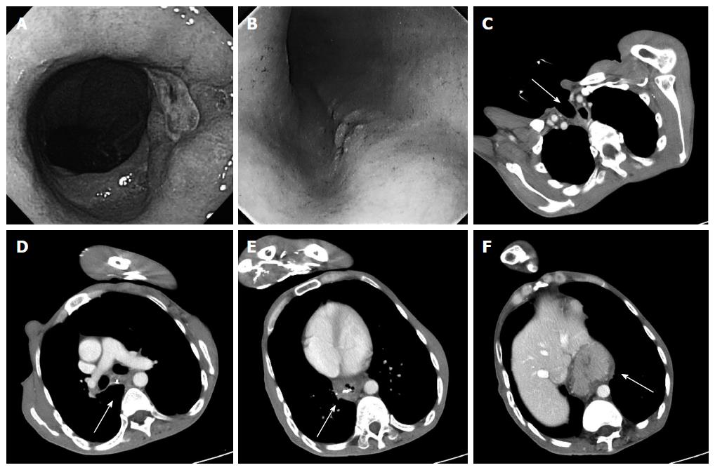
Figure 1 Endoscopy findings (A, B) and computed tomography findings (C-F).
A: An ulcerative lesion was found in a hiatal hernia 30 cm from an incisor; B: 0-IIc type esophageal cancer was detected 25 cm from an incisor; There was severe deformity of the trunk caused by kyphosis and muscular contractures (C-F); C: The arrow points to the permanent tracheal stoma; D: The arrow points to the metal clip near the 0-IIc type esophageal cancer; E: The arrow points to the metal clip near the ulcerative lesion in hiatal hernia; F: The arrow points to the sliding hiatal hernia.
- Citation: Shiozaki A, Fujiwara H, Konishi H, Kinoshita O, Kosuga T, Morimura R, Murayama Y, Komatsu S, Kuriu Y, Ikoma H, Nakanishi M, Ichikawa D, Okamoto K, Sakakura C, Otsuji E. Laparoscopic transhiatal approach for resection of an adenocarcinoma in long-segment Barrett’s esophagus. World J Gastroenterol 2015; 21(29): 8974-8980
- URL: https://www.wjgnet.com/1007-9327/full/v21/i29/8974.htm
- DOI: https://dx.doi.org/10.3748/wjg.v21.i29.8974


