Copyright
©The Author(s) 2015.
World J Gastroenterol. Aug 7, 2015; 21(29): 8878-8887
Published online Aug 7, 2015. doi: 10.3748/wjg.v21.i29.8878
Published online Aug 7, 2015. doi: 10.3748/wjg.v21.i29.8878
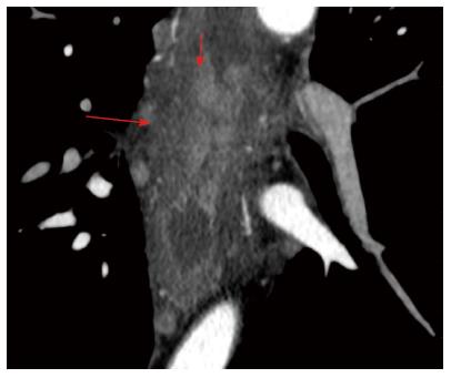
Figure 7 Atypical manifestation of corrosive esophageal stricture at computed tomography imaging.
Arterial phase. Axial computed tomography (CT) scan of corrosive esophageal stricture. Development of mucosa granulations (short arrow), fibrotically changed submucosal and muscular layers and adventitia of esophageal walls (long arrow).
- Citation: Karmazanovsky GG, Buryakina SA, Kondratiev EV, Yang Q, Ruchkin DV, Kalinin DV. Value of two-phase dynamic multidetector computed tomography in differential diagnosis of post-inflammatory strictures from esophageal cancer. World J Gastroenterol 2015; 21(29): 8878-8887
- URL: https://www.wjgnet.com/1007-9327/full/v21/i29/8878.htm
- DOI: https://dx.doi.org/10.3748/wjg.v21.i29.8878


