Copyright
©The Author(s) 2015.
World J Gastroenterol. Aug 7, 2015; 21(29): 8878-8887
Published online Aug 7, 2015. doi: 10.3748/wjg.v21.i29.8878
Published online Aug 7, 2015. doi: 10.3748/wjg.v21.i29.8878
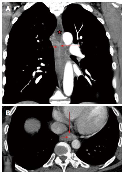
Figure 2 Corrosive esophageal stricture.
A: Multidetector computed tomography (MDCT). Arterial phase. Coronal reconstruction. Concentric esophageal wall thickening of the corrosive stricture, which has a homogeneous structure (short arrow). The mucosa is traceable as a thin hyperintense line in the centre of thickened walls that was caused by fibrotic changes (long arrow). Conically shaped suprastenotic dilatation (star) with smooth upper boundaries of the stricture; B: MDCT. Arterial phase. Axial CT scan. Target sign - thickening of saved esophageal mucosa (short arrow) in the centre of fibrotically changed submucosal, muscular layers and adventitia of esophageal walls (long arrow).
- Citation: Karmazanovsky GG, Buryakina SA, Kondratiev EV, Yang Q, Ruchkin DV, Kalinin DV. Value of two-phase dynamic multidetector computed tomography in differential diagnosis of post-inflammatory strictures from esophageal cancer. World J Gastroenterol 2015; 21(29): 8878-8887
- URL: https://www.wjgnet.com/1007-9327/full/v21/i29/8878.htm
- DOI: https://dx.doi.org/10.3748/wjg.v21.i29.8878


