Copyright
©The Author(s) 2015.
World J Gastroenterol. Aug 7, 2015; 21(29): 8848-8857
Published online Aug 7, 2015. doi: 10.3748/wjg.v21.i29.8848
Published online Aug 7, 2015. doi: 10.3748/wjg.v21.i29.8848
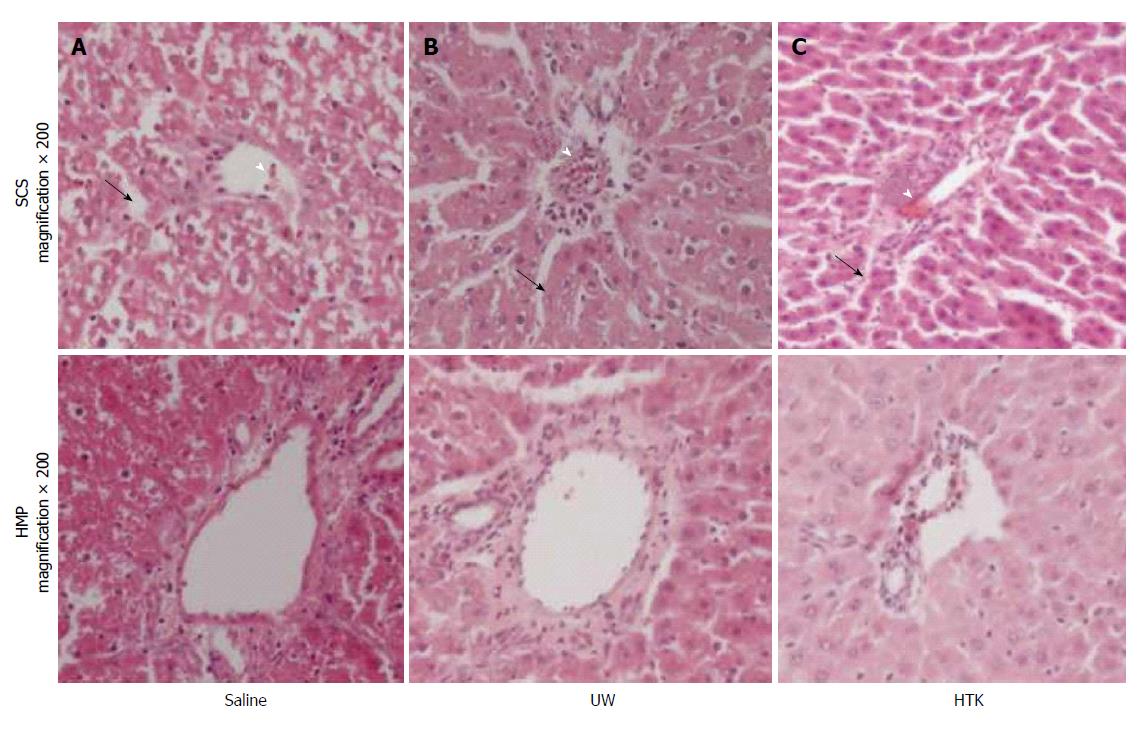
Figure 2 Histopathological appearance of the livers in the studies at 6 h after perfusion (hematoxylin and eosin staining, original magnification × 200).
A: Saline; B: UW; C: HTK. Sinusoid lining (arrow) and congestion of the sinusoids and central vein (white arrow head).
- Citation: Jia JJ, Zhang J, Li JH, Chen XD, Jiang L, Zhou YF, He N, Xie HY, Zhou L, Zheng SS. Influence of perfusate on liver viability during hypothermic machine perfusion. World J Gastroenterol 2015; 21(29): 8848-8857
- URL: https://www.wjgnet.com/1007-9327/full/v21/i29/8848.htm
- DOI: https://dx.doi.org/10.3748/wjg.v21.i29.8848


