Copyright
©The Author(s) 2015.
World J Gastroenterol. Aug 7, 2015; 21(29): 8739-8752
Published online Aug 7, 2015. doi: 10.3748/wjg.v21.i29.8739
Published online Aug 7, 2015. doi: 10.3748/wjg.v21.i29.8739
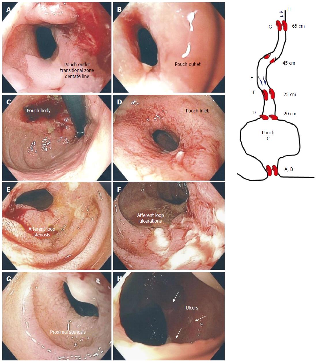
Figure 5 Crohn’s ileo-pouchitis.
Stenosis of the pouch outlet (A, B), normal appearing mucosa of the pouch (C), stenosis of the pouch inlet (D), stenosis of the afferent limb at 25 cm (E), linear deep ulcerations in the ileum (F), proximal stenosis of the ileum at 65 cm (G) and ulcers more proximally (H).
- Citation: Zezos P, Saibil F. Inflammatory pouch disease: The spectrum of pouchitis. World J Gastroenterol 2015; 21(29): 8739-8752
- URL: https://www.wjgnet.com/1007-9327/full/v21/i29/8739.htm
- DOI: https://dx.doi.org/10.3748/wjg.v21.i29.8739


