Copyright
©The Author(s) 2015.
World J Gastroenterol. Jul 28, 2015; 21(28): 8730-8738
Published online Jul 28, 2015. doi: 10.3748/wjg.v21.i28.8730
Published online Jul 28, 2015. doi: 10.3748/wjg.v21.i28.8730
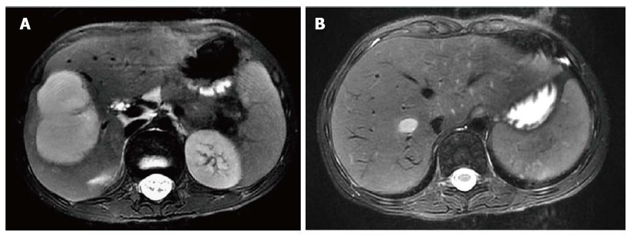
Figure 2 T2-attenuated magnetic resonance imaging scan of the abdomen.
A: A 4.7 cm × 4.7 cm × 6.6 cm, contrast-enhancing, hyper-intensive, well-defined, and moderate- to large-sized lesion involving the right hepatic lobe (segments# V, VI and VII); B: There was extension of the known hepatic IPT lesion into the path of the right hepatic vein.
- Citation: Al-Hussaini H, Azouz H, Abu-Zaid A. Hepatic inflammatory pseudotumor presenting in an 8-year-old boy: A case report and review of literature. World J Gastroenterol 2015; 21(28): 8730-8738
- URL: https://www.wjgnet.com/1007-9327/full/v21/i28/8730.htm
- DOI: https://dx.doi.org/10.3748/wjg.v21.i28.8730


