Copyright
©The Author(s) 2015.
World J Gastroenterol. Jul 28, 2015; 21(28): 8723-8729
Published online Jul 28, 2015. doi: 10.3748/wjg.v21.i28.8723
Published online Jul 28, 2015. doi: 10.3748/wjg.v21.i28.8723
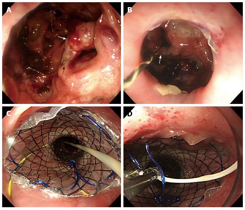
Figure 3 Endoscopic findings of gastric conduit necrosis and self-expanding metal stent placement.
A: The stump of the healthy gastric conduit (thick white arrows) and the staple in the stump of the gastric conduit (thin black arrow); B: Complete disruption of 2 cm of the anastomosis and stump of the gastric conduit (thick white arrows); C: Placement of a removable covered self-expanding metal stent (SEMS); D: A silk thread placed through the lasso located on the proximal end of the stent.
- Citation: Oshikiri T, Yamamoto Y, Miki I, Tsuda M, Nakamura T, Fujino Y, Tominaga M, Kakeji Y. Conservative reconstruction using stents as salvage therapy for disruption of esophago-gastric anastomosis. World J Gastroenterol 2015; 21(28): 8723-8729
- URL: https://www.wjgnet.com/1007-9327/full/v21/i28/8723.htm
- DOI: https://dx.doi.org/10.3748/wjg.v21.i28.8723


