Copyright
©The Author(s) 2015.
World J Gastroenterol. Jul 21, 2015; 21(27): 8467-8472
Published online Jul 21, 2015. doi: 10.3748/wjg.v21.i27.8467
Published online Jul 21, 2015. doi: 10.3748/wjg.v21.i27.8467
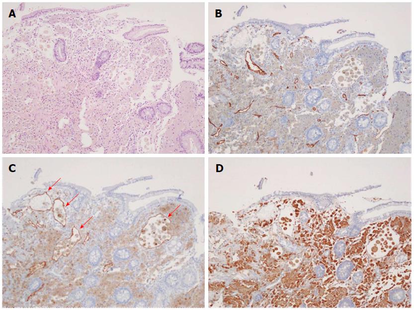
Figure 5 Histological examination of the terminal biopsy specimens.
A: Hematoxylin and eosin stain (magnification × 100) showing dilated lymphatic vessels with many foamy macrophages in the lamina propria consistent with lymphangiectasia; B: CD34 stain showing normal vascular endothelial cells; C: D2-40 stained endothelial cells (red arrows), indicating dilated lymphatics; D: CD68-stained macrophages, which have aggregated to uptake lipids leaking from dilated lymphatics.
- Citation: Lee SJ, Song HJ, Boo SJ, Na SY, Kim HU, Hyun CL. Primary intestinal lymphangiectasia with generalized warts. World J Gastroenterol 2015; 21(27): 8467-8472
- URL: https://www.wjgnet.com/1007-9327/full/v21/i27/8467.htm
- DOI: https://dx.doi.org/10.3748/wjg.v21.i27.8467


