Copyright
©The Author(s) 2015.
World J Gastroenterol. Jul 21, 2015; 21(27): 8452-8457
Published online Jul 21, 2015. doi: 10.3748/wjg.v21.i27.8452
Published online Jul 21, 2015. doi: 10.3748/wjg.v21.i27.8452
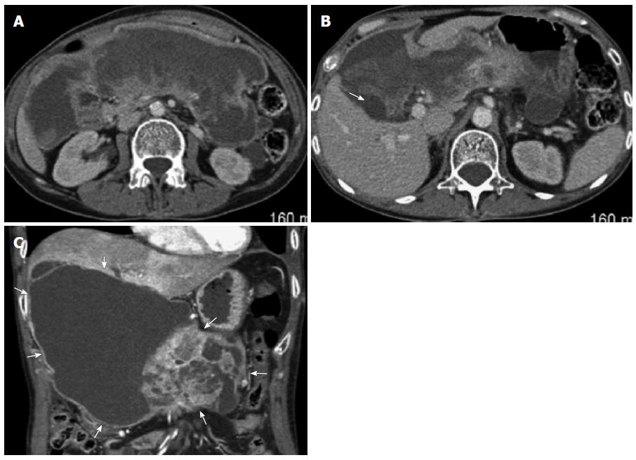
Figure 3 Post-contrast venous phase abdominal computed tomography.
A: Demonstrating a large (20 cm × 6 cm × 12 cm) abdominal mass with peripheral rim enhancement as well as some areas of thickened nodular enhancement in the periphery. There is a wispy enhancing area of probable pancreatic tissue in the posterior mid aspect of the mass in the expected location of the pancreatic neck, body and proximal tail. The portal splenic confluence and downstream SMV are inseparable from the mass; B: The cystic mass abuts the liver at the gallbladder fossa, and surrounds the gallbladder (arrow); C: Coronal reconstruction. Giant cystic-predominant mass (arrows) extends from the hepatic hilum, and surrounds the gallbladder.
- Citation: Akpinar B, Obuch J, Fukami N, Pokharel SS. Unusual presentation of a pancreatic cyst resulting from osteosarcoma metastasis. World J Gastroenterol 2015; 21(27): 8452-8457
- URL: https://www.wjgnet.com/1007-9327/full/v21/i27/8452.htm
- DOI: https://dx.doi.org/10.3748/wjg.v21.i27.8452


