Copyright
©The Author(s) 2015.
World J Gastroenterol. Jul 21, 2015; 21(27): 8452-8457
Published online Jul 21, 2015. doi: 10.3748/wjg.v21.i27.8452
Published online Jul 21, 2015. doi: 10.3748/wjg.v21.i27.8452
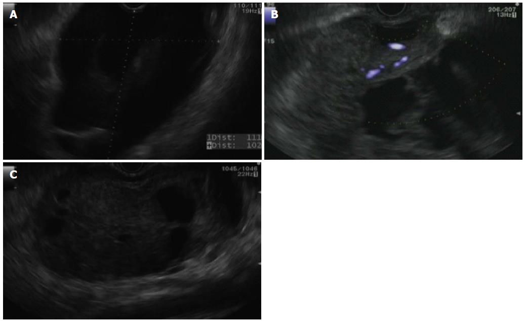
Figure 2 Endoscopic ultrasound imaging.
A: Showing a large pancreatic cystic mass in the head/genu of the pancreas; B: Showing blood flow within solid components of the pancreatic mass (blue Doppler flow); C: Showing decompression of the pancreatic cystic lesion with residual solid component.
- Citation: Akpinar B, Obuch J, Fukami N, Pokharel SS. Unusual presentation of a pancreatic cyst resulting from osteosarcoma metastasis. World J Gastroenterol 2015; 21(27): 8452-8457
- URL: https://www.wjgnet.com/1007-9327/full/v21/i27/8452.htm
- DOI: https://dx.doi.org/10.3748/wjg.v21.i27.8452


