Copyright
©The Author(s) 2015.
World J Gastroenterol. Jul 14, 2015; 21(26): 8215-8220
Published online Jul 14, 2015. doi: 10.3748/wjg.v21.i26.8215
Published online Jul 14, 2015. doi: 10.3748/wjg.v21.i26.8215
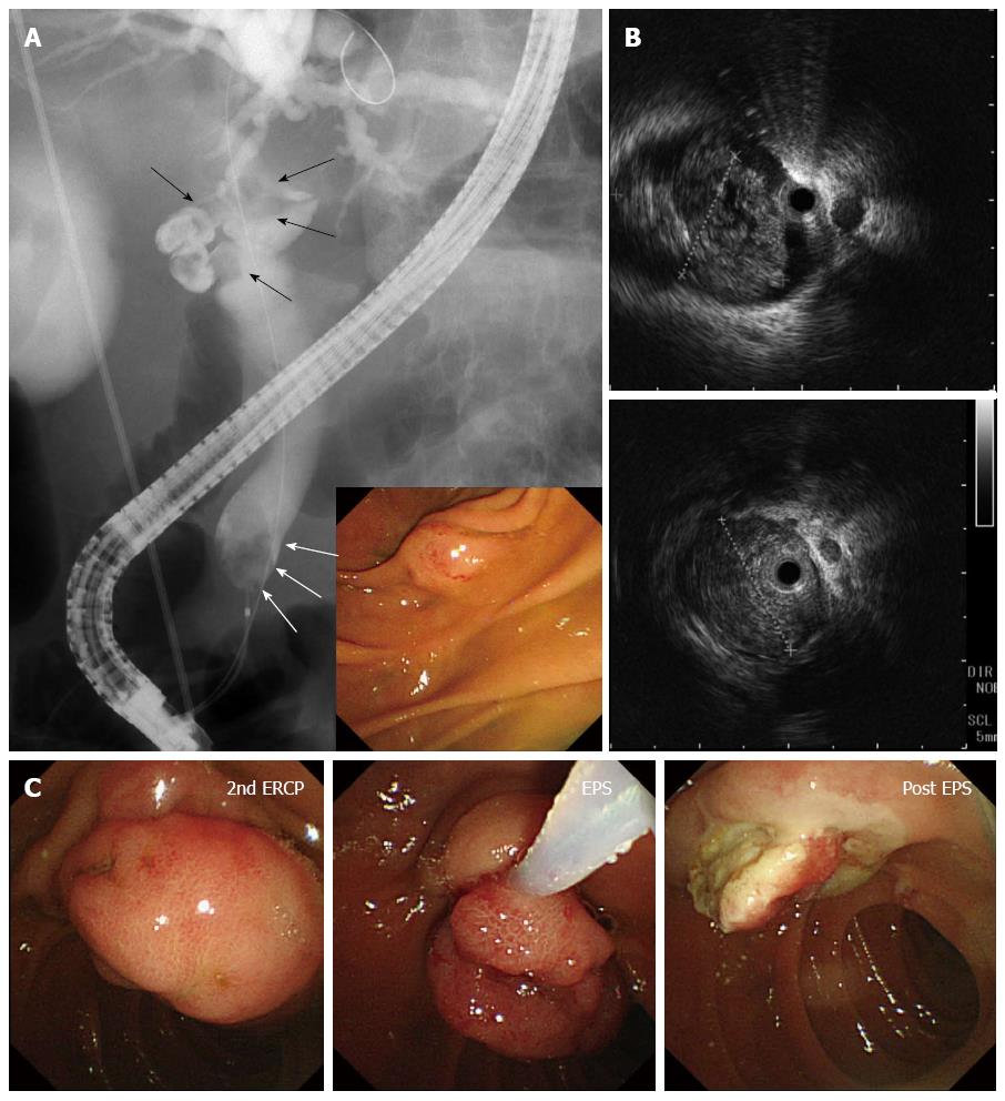
Figure 3 Tumor resection by endoscopic snare papillectomy.
A: Endoscopic retrograde cholangiopancreatography (ERCP) showing several filling defects in the upper part of the common bile duct (CBD) (black arrows), and an oval-shaped defect in the intra-pancreatic CBD (white arrows). Endoscopic picture during ERCP did not show any remarkable findings without juxtapapillary duodenal diverticula; B: Intraductal ultrasonography confirming that the lower filling defect was a papillary-growing tumor in the CBD; C: Second ERCP, performed one week later, showing inversion and exclusion of the tumor after endoscopic sphincterotomy. The tumor was a pedunculated polyp, enabling en bloc resection by endoscopic snare papillectomy.
- Citation: Suzuki K, Higuchi H, Shimizu S, Nakano M, Serizawa H, Morinaga S. Endoscopic snare papillectomy for a solitary Peutz-Jeghers-type polyp in the duodenum with ingrowth into the common bile duct: Case report. World J Gastroenterol 2015; 21(26): 8215-8220
- URL: https://www.wjgnet.com/1007-9327/full/v21/i26/8215.htm
- DOI: https://dx.doi.org/10.3748/wjg.v21.i26.8215


