Copyright
©The Author(s) 2015.
World J Gastroenterol. Jul 14, 2015; 21(26): 8208-8214
Published online Jul 14, 2015. doi: 10.3748/wjg.v21.i26.8208
Published online Jul 14, 2015. doi: 10.3748/wjg.v21.i26.8208
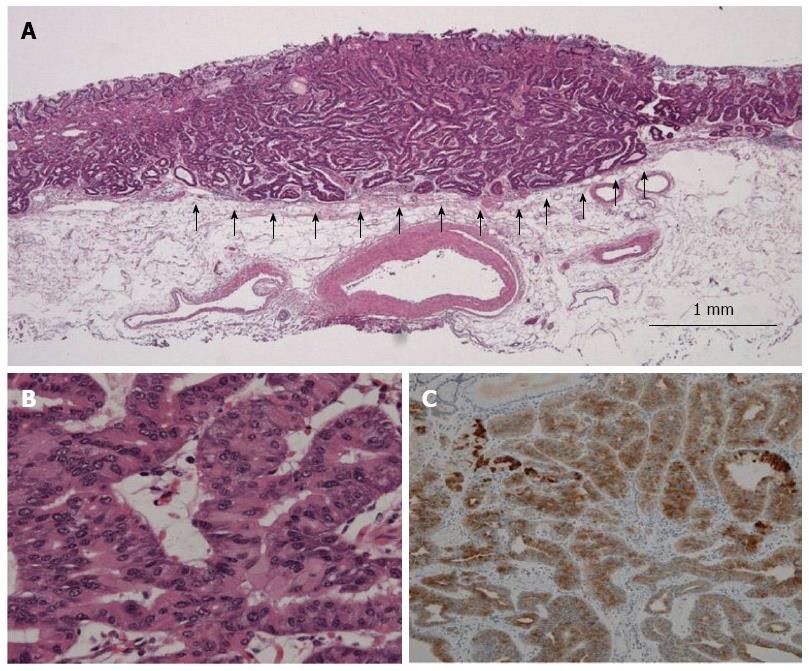
Figure 2 Pathological examination of resected gastric adenocarcinoma of fundic gland type (case 1).
A: In low-power view, the tumor arose from the deep layer of the lamina propria mucosae and invaded the submucosal layer at a depth of 980 μm (arrow). Most of the surface was covered with non-atypical foveolar epithelium; B: In high-power view, the tumor was composed of well-differentiated columnar cells mimicking the fundic gland cells with mild nuclear atypism; C: Immunohistological staining showed diffuse positivity for pepsinogen-I.
- Citation: Miyazawa M, Matsuda M, Yano M, Hara Y, Arihara F, Horita Y, Matsuda K, Sakai A, Noda Y. Gastric adenocarcinoma of fundic gland type: Five cases treated with endoscopic resection. World J Gastroenterol 2015; 21(26): 8208-8214
- URL: https://www.wjgnet.com/1007-9327/full/v21/i26/8208.htm
- DOI: https://dx.doi.org/10.3748/wjg.v21.i26.8208


