Copyright
©The Author(s) 2015.
World J Gastroenterol. Jul 14, 2015; 21(26): 8073-8080
Published online Jul 14, 2015. doi: 10.3748/wjg.v21.i26.8073
Published online Jul 14, 2015. doi: 10.3748/wjg.v21.i26.8073
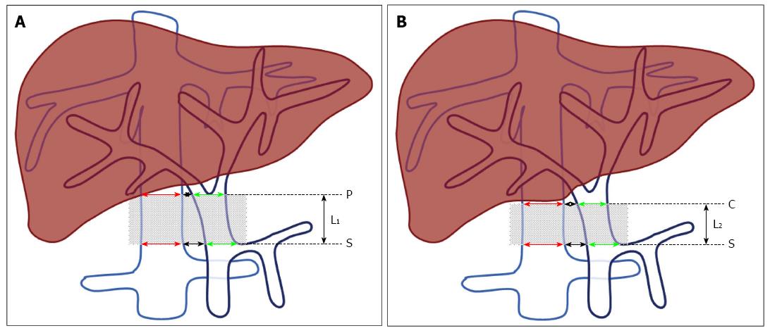
Figure 1 Effective length of the portacaval anastomosis using the magnetic compression technique.
A: When the origin of the portal vein (PV) branch is below the lower edge of the caudate lobe, the effective length, L1, is the distance between the intersection of the splenic vein and the PV, here S, and the origin of the PV branching, P. B: When the origin of the PV branch is higher than the lower edge of the caudate lobe, the effective length, L2, is the distance between S and the lower edge of caudate lobe, C. The red arrow indicates the diameter of the inferior vena cava (IVC); the green arrow indicates the diameter of the PV; the black arrow indicates the distance between the PV and the IVC.
-
Citation: Yan XP, Liu WY, Ma J, Li JP, Lv Y. Extrahepatic portacaval shunt
via a magnetic compression technique: A cadaveric feasibility study. World J Gastroenterol 2015; 21(26): 8073-8080 - URL: https://www.wjgnet.com/1007-9327/full/v21/i26/8073.htm
- DOI: https://dx.doi.org/10.3748/wjg.v21.i26.8073


