Copyright
©The Author(s) 2015.
World J Gastroenterol. Jul 14, 2015; 21(26): 8043-8051
Published online Jul 14, 2015. doi: 10.3748/wjg.v21.i26.8043
Published online Jul 14, 2015. doi: 10.3748/wjg.v21.i26.8043
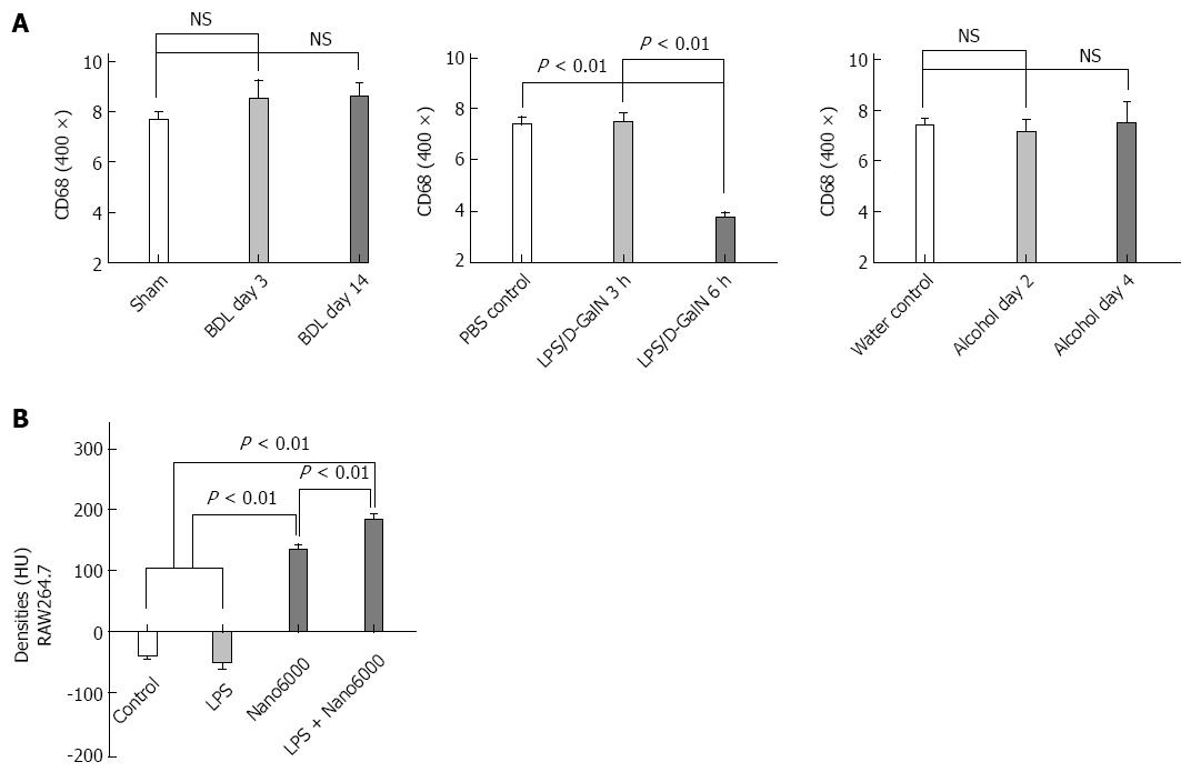
Figure 5 Comparison of the number of CD68+ cells in the injured livers of the three models (A); comparison of the densities of RAW264.
7 cell mass co-cultured with nano6000, lipopolysaccharide or both (B). Values are represented as the means of triplicate values and presented as the mean ± SD.
- Citation: Hua XW, Lu TF, Li DW, Wang WG, Li J, Liu ZZ, Lin WW, Zhang JJ, Xia Q. Contrast-enhanced micro-computed tomography using ExiTron nano6000 for assessment of liver injury. World J Gastroenterol 2015; 21(26): 8043-8051
- URL: https://www.wjgnet.com/1007-9327/full/v21/i26/8043.htm
- DOI: https://dx.doi.org/10.3748/wjg.v21.i26.8043


