Copyright
©The Author(s) 2015.
World J Gastroenterol. Jul 7, 2015; 21(25): 7824-7833
Published online Jul 7, 2015. doi: 10.3748/wjg.v21.i25.7824
Published online Jul 7, 2015. doi: 10.3748/wjg.v21.i25.7824
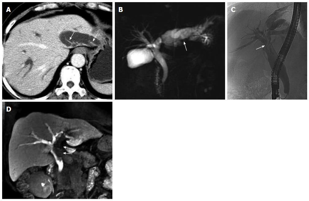
Figure 1 Fifty-nine-year-old female patient with intraductal papillary mucinous neoplasm of the bile duct (case 1).
A: Axial contrast-enhanced computed tomography in the portal phase; B: Magnetic resonance cholangiopancreatography showed extensive intra- and extrahepatic bile duct dilatation and multiple hydra-like protrusions (arrows) in the markedly dilated left intrahepatic bile duct (arrow); C: Endoscopic retrograde cholangiopancreatography showed extensive intra- and extrahepatic bile duct dilatation, particularly in the left intrahepatic and common bile ducts. Bile ducts in the liver hilar appeared more transparent due to the local aggregation of mucin (arrow); D: Multiple planar reconstruction of gadolinium-ethoxybenzyl-diethylenetriamine-pentaacetic acid-enhanced magnetic resonance imaging in the hepatobiliary phase showed dilatation of the bile duct and filling defects in the left intrahepatic and common bile ducts, with a cup-shaped contrast filling edge (arrows). The filling defects were caused by mucus retention, which was confirmed by endoscopy.
- Citation: Ying SH, Teng XD, Wang ZM, Wang QD, Zhao YL, Chen F, Xiao WB. Gd-EOB-DTPA-enhanced magnetic resonance imaging for bile duct intraductal papillary mucinous neoplasms. World J Gastroenterol 2015; 21(25): 7824-7833
- URL: https://www.wjgnet.com/1007-9327/full/v21/i25/7824.htm
- DOI: https://dx.doi.org/10.3748/wjg.v21.i25.7824


