Copyright
©The Author(s) 2015.
World J Gastroenterol. Jun 28, 2015; 21(24): 7608-7612
Published online Jun 28, 2015. doi: 10.3748/wjg.v21.i24.7608
Published online Jun 28, 2015. doi: 10.3748/wjg.v21.i24.7608
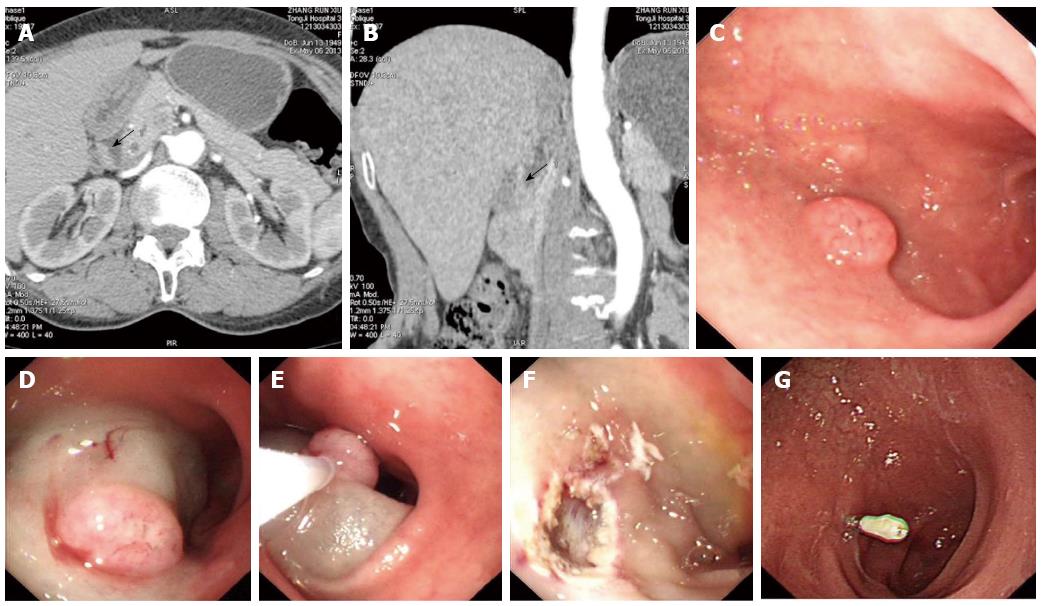
Figure 1 Computed tomography, endoscopic image and endoscopic mucosal resection of duodenal tumor with neuroendocrine features.
A: Computed tomography (CT) image of a duodenal tumor (arrow); B: Three dimensional CT image reconstruction of the duodenal tumor (arrow); C: Endoscopic image of the duodenal tumor; D: Positive lifting sign after injection of physiological saline with 1:10000 norepinephrine; E: Endoscopic mucosal resection after snare; F: Clean border of the wound after endoscopic mucosal resection (EMR); G: Scar formation on EMR position at gastroscopic review after two months.
- Citation: Wen MY, Wang Y, Meng XY, Xie HP. Endoscopic mucosal resection of duodenal bulb adenocarcinoma with neuroendocrine features: An extremely rare case report. World J Gastroenterol 2015; 21(24): 7608-7612
- URL: https://www.wjgnet.com/1007-9327/full/v21/i24/7608.htm
- DOI: https://dx.doi.org/10.3748/wjg.v21.i24.7608


