Copyright
©The Author(s) 2015.
World J Gastroenterol. Jun 28, 2015; 21(24): 7604-7607
Published online Jun 28, 2015. doi: 10.3748/wjg.v21.i24.7604
Published online Jun 28, 2015. doi: 10.3748/wjg.v21.i24.7604
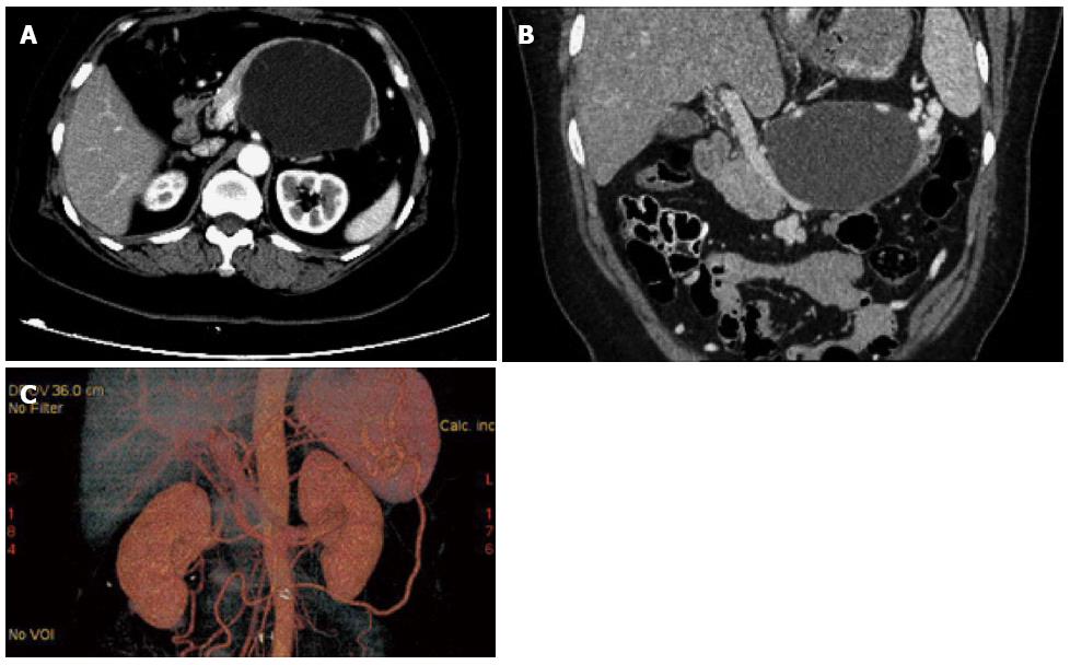
Figure 1 Preoperative computed tomography scan.
A: Abdominal contrast-enhanced computed tomography (CT) showed a cystic tumor in the body-tail of the pancreas; B: Coronal multiplanar reformation showed that the lesion was adhered to and constricted the main trunk of the superior mesenteric vein; C: 3-dimensional CT scan showed the presence of several well-developed collateral vessels.
- Citation: Chen YT, Jiang QL, Zhu Z, Wang S, Zhao XM, Lan ZM, Che X, Zhang JW, Cui L, Tang XL, Wang CF. Resection of the main trunk of the superior mesenteric vein without reconstruction during surgery for giant pancreatic mucinous cystadenoma: A case report. World J Gastroenterol 2015; 21(24): 7604-7607
- URL: https://www.wjgnet.com/1007-9327/full/v21/i24/7604.htm
- DOI: https://dx.doi.org/10.3748/wjg.v21.i24.7604


