Copyright
©The Author(s) 2015.
World J Gastroenterol. Jun 28, 2015; 21(24): 7598-7603
Published online Jun 28, 2015. doi: 10.3748/wjg.v21.i24.7598
Published online Jun 28, 2015. doi: 10.3748/wjg.v21.i24.7598
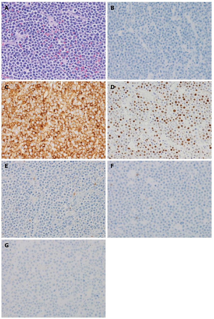
Figure 3 Plasmablastic lymphoma of the sigmoid colon.
A: Tumor cells have large hyperchromatic nuclei with prominent nucleoli. Some tumor cells showed plasmacytic differentiation [hematoxylin and eosin (HE) staining, original magnification × 400]; B: The atypical cells are negative for the B-cell marker CD20 (original magnification × 400); C: The atypical cells are diffusely positive for the plasma cell marker CD138 (original magnification × 400); D: The nuclei of the atypical cells are positive for EBER, which is Epstein-Barr encoded RNA (original magnification × 400); E: The atypical cells are negative for the leukocyte common antigen CD45RO (UCHL1) (original magnification × 400); F: The atypical cells are negative for the T-cell marker CD3 (original magnification × 400); G: The atypical cells are negative for the pan-B-cell marker CD79a (original magnification × 400).
- Citation: Haramura T, Haraguchi M, Irie J, Ito S, Tokai H, Noda K, Kitajima M, Minami S, Inoue K, Sasaki Y, Oshima K, Eguchi S. Case of plasmablastic lymphoma of the sigmoid colon and literature review. World J Gastroenterol 2015; 21(24): 7598-7603
- URL: https://www.wjgnet.com/1007-9327/full/v21/i24/7598.htm
- DOI: https://dx.doi.org/10.3748/wjg.v21.i24.7598


