Copyright
©The Author(s) 2015.
World J Gastroenterol. Jun 28, 2015; 21(24): 7589-7593
Published online Jun 28, 2015. doi: 10.3748/wjg.v21.i24.7589
Published online Jun 28, 2015. doi: 10.3748/wjg.v21.i24.7589
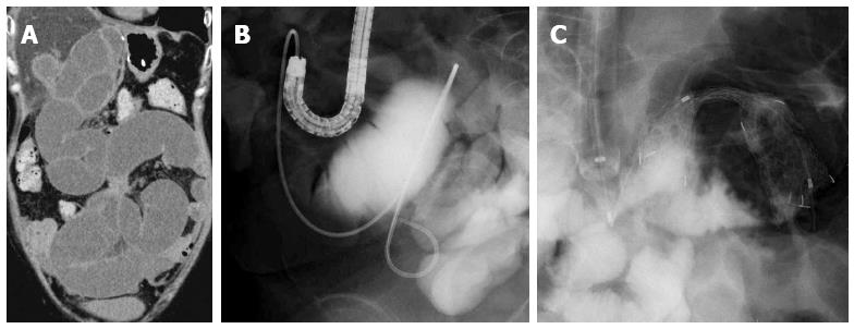
Figure 3 Computed tomography shows dilatation of intestine due to peritoneum dissemination (A); an endoscopic nasal drainage tube was placed into the dilated intestine through the endoscopic working channel (B) and an enteral metallic stent was placed through the stenosis along the guidewire in the overtube (C).
- Citation: Nakahara K, Okuse C, Matsumoto N, Suetani K, Morita R, Michikawa Y, Ozawa SI, Hosoya K, Kobayashi S, Otsubo T, Itoh F. Enteral metallic stenting by balloon enteroscopy for obstruction of surgically reconstructed intestine. World J Gastroenterol 2015; 21(24): 7589-7593
- URL: https://www.wjgnet.com/1007-9327/full/v21/i24/7589.htm
- DOI: https://dx.doi.org/10.3748/wjg.v21.i24.7589


