Copyright
©The Author(s) 2015.
World J Gastroenterol. Jun 28, 2015; 21(24): 7457-7467
Published online Jun 28, 2015. doi: 10.3748/wjg.v21.i24.7457
Published online Jun 28, 2015. doi: 10.3748/wjg.v21.i24.7457
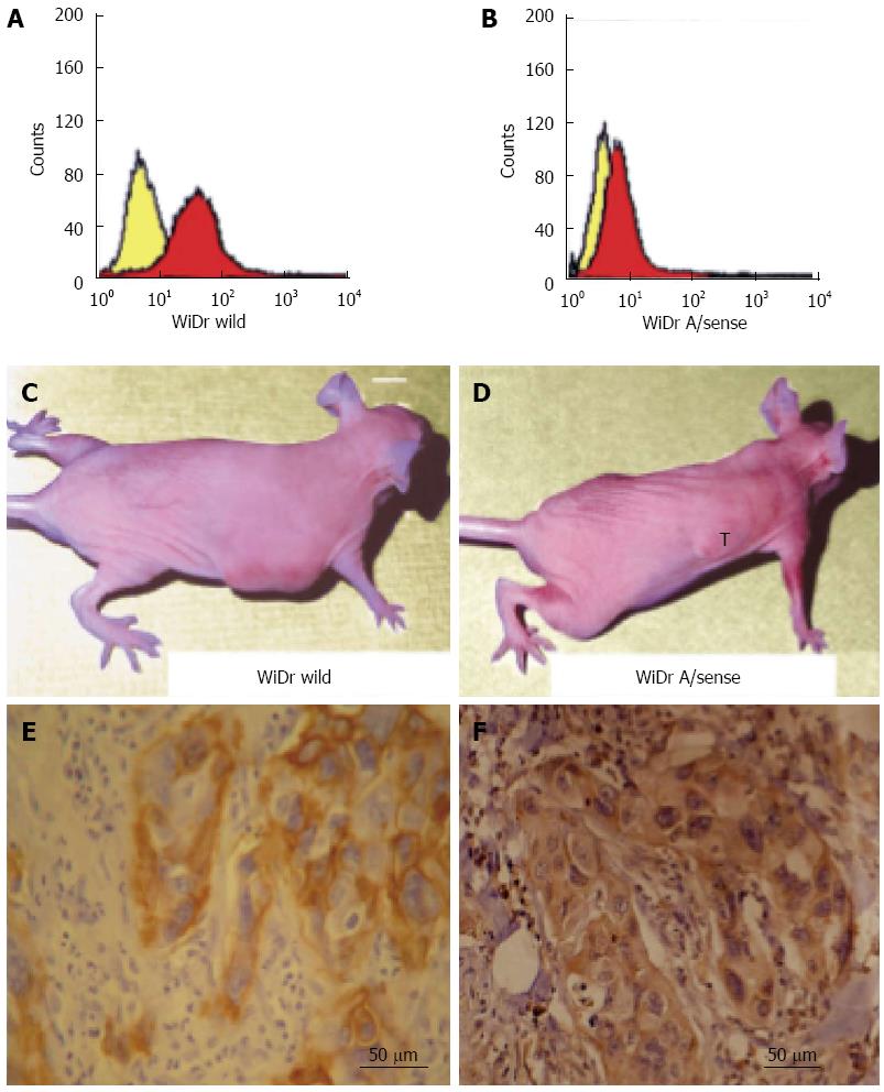
Figure 4 Integrin αvβ6 promotes invasive tumor growth in nude mice.
Wild-type WiDr cells (A) and antisense β6 WiDr transfectants (B) were analyzed by FACScan for the expression of integrin αvβ6. The yellow and red histograms represent analyses in the absence and presence of E7P6 primary antibody; the secondary antibody (goat anti-mouse IgG) was conjugated with phycoerythrin. Tumor invasive growth after 6 wk following subcutaneous inoculation with wild-type WiDr cells (C) or antisense beta 6 WiDr transfectants (D). Increased expression of integrin αvβ6 (E) and matrix metalloproteinase-9 (MMP-9) (F) was identified in serial sections of tumor xenografts inoculated with wild-type WiDr cells. One section was selected from every 10 serial sections. The figures (E) and (F) are the 1st section and 21st section selected. The thickness of each section was 5 μm. A scale bar is shown in the right lower corner of (E) and (F).
- Citation: Yang GY, Guo S, Dong CY, Wang XQ, Hu BY, Liu YF, Chen YW, Niu J, Dong JH. Integrin αvβ6 sustains and promotes tumor invasive growth in colon cancer progression. World J Gastroenterol 2015; 21(24): 7457-7467
- URL: https://www.wjgnet.com/1007-9327/full/v21/i24/7457.htm
- DOI: https://dx.doi.org/10.3748/wjg.v21.i24.7457


