Copyright
©The Author(s) 2015.
World J Gastroenterol. Jun 21, 2015; 21(23): 7335-7342
Published online Jun 21, 2015. doi: 10.3748/wjg.v21.i23.7335
Published online Jun 21, 2015. doi: 10.3748/wjg.v21.i23.7335
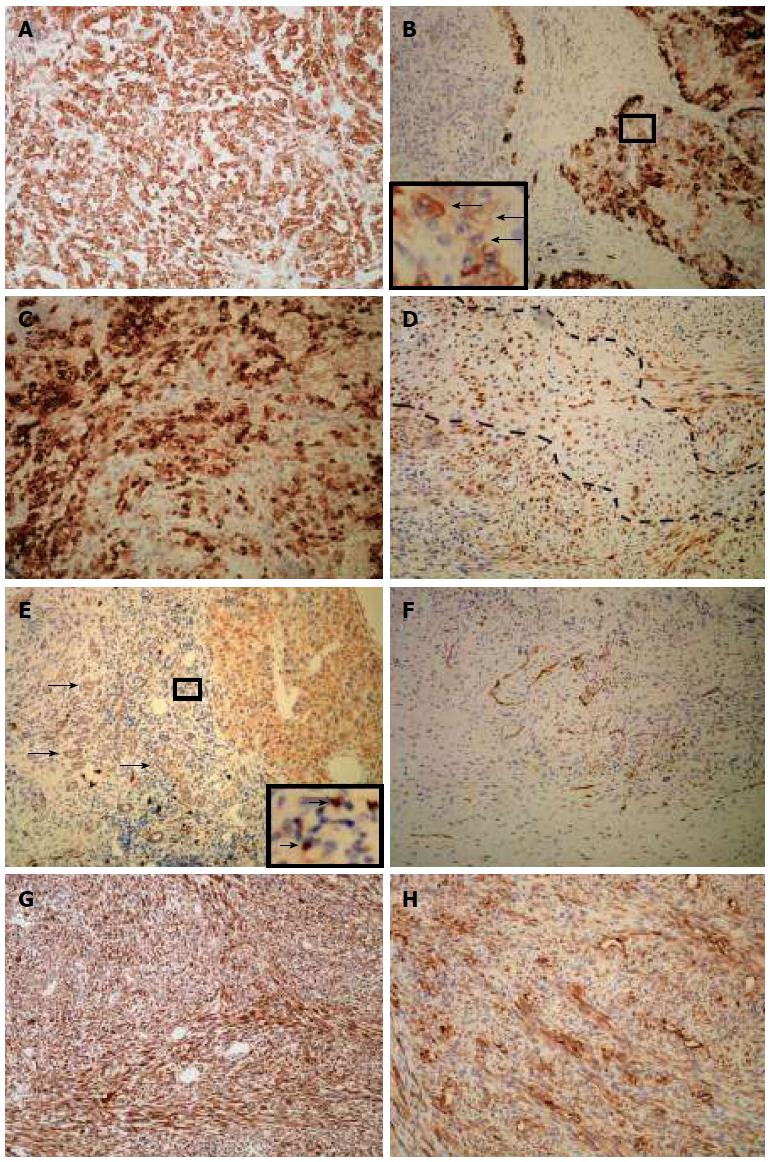
Figure 3 Immunohistochemical expression of the carcinosarcoma.
A: Pan-cytokeratin staining showed diffuse and strong signals for HCC cells; B: HCC elements showed various staining pattern for cytokeratin 19 with intermediate hepatobiliary cell-like tumor cells (arrows); C: Carcinomatous elements were strongly positive for epithelial membrane antigen; D: Spindle cells and chondrosarcoma cells (area fenced by dotted line) were positive for epithelial membrane antigen; E: Carcinoma cells were positive for CD117, as were ductular reactions (arrows) and undifferentiated cells (arrows, inset); F: Sarcoma cells were positive for CD117; G: Sarcomatous elements strongly stained for vimentin; H: Diffuse staining for smooth muscle actin in sarcoma cells; All magnifications are × 100.
- Citation: Xiang S, Chen YF, Guan Y, Chen XP. Primary combined hepatocellular-cholangiocellular sarcoma: An unusual case. World J Gastroenterol 2015; 21(23): 7335-7342
- URL: https://www.wjgnet.com/1007-9327/full/v21/i23/7335.htm
- DOI: https://dx.doi.org/10.3748/wjg.v21.i23.7335


