Copyright
©The Author(s) 2015.
World J Gastroenterol. Jun 21, 2015; 21(23): 7326-7330
Published online Jun 21, 2015. doi: 10.3748/wjg.v21.i23.7326
Published online Jun 21, 2015. doi: 10.3748/wjg.v21.i23.7326
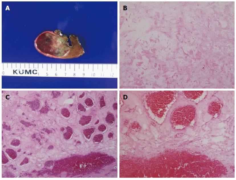
Figure 3 Gross photographic and pathological findings.
A: Macroscopic specimen showing intratumoral hemorrhage within hemangioma; B: Variably sized vascular spaces lined with flat endothelial cells and myxoid stroma [hematoxylin-eosin (HE) staining, magnification × 40]; C: Widely dilated vascular spaces filled with thrombi in the resected specimen (HE staining, magnification × 40); D: HE staining, magnification × 100.
- Citation: Kim JM, Chung WJ, Jang BK, Hwang JS, Kim YH, Kwon JH, Choi MS. Hemorrhagic hemangioma in the liver: A case report. World J Gastroenterol 2015; 21(23): 7326-7330
- URL: https://www.wjgnet.com/1007-9327/full/v21/i23/7326.htm
- DOI: https://dx.doi.org/10.3748/wjg.v21.i23.7326


