Copyright
©The Author(s) 2015.
World J Gastroenterol. Jun 21, 2015; 21(23): 7225-7232
Published online Jun 21, 2015. doi: 10.3748/wjg.v21.i23.7225
Published online Jun 21, 2015. doi: 10.3748/wjg.v21.i23.7225
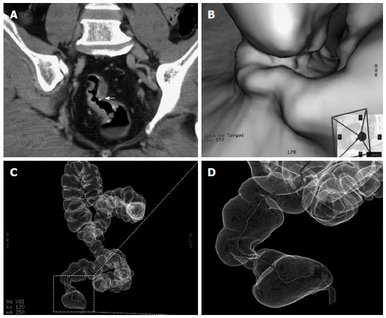
Figure 3 Computed tomography presentation.
A: Computed tomography (CT) scan showing curvilinear calcification in the rectosigmoid colon and calcified, conglomerate nodules (arrow) protruding from the wall of the rectosigmoid colon; B: Lobulated polypus in the rectum; C, D: CTVC enables three-dimensional view of walls of the colon as a result of reconstruction of multislice CT images. The colorectal stenosis is showed in the area surrounding by dotted lines. CTVC: CT virtual colonoscopy.
- Citation: Feng H, Lu AG, Zhao XW, Han DP, Zhao JK, Shi L, Schiergens TS, Lee SM, Zhang WP, Thasler WE. Comparison of non-schistosomal rectosigmoid cancer and schistosomal rectosigmoid cancer. World J Gastroenterol 2015; 21(23): 7225-7232
- URL: https://www.wjgnet.com/1007-9327/full/v21/i23/7225.htm
- DOI: https://dx.doi.org/10.3748/wjg.v21.i23.7225


