Copyright
©The Author(s) 2015.
World J Gastroenterol. Jun 21, 2015; 21(23): 7172-7180
Published online Jun 21, 2015. doi: 10.3748/wjg.v21.i23.7172
Published online Jun 21, 2015. doi: 10.3748/wjg.v21.i23.7172
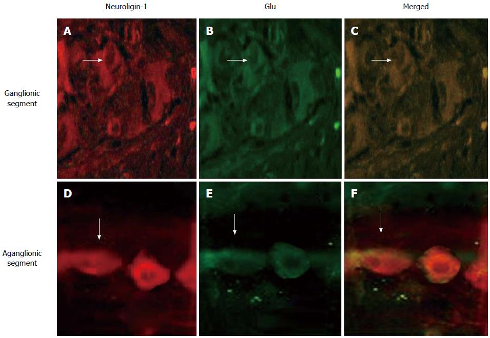
Figure 1 Both in ganglionic segment and aganglionic segment, neuroligin-1 (A, D, red) is expressed in the same position (merged, C, F, yellow) where glutamate is expressed (B, E, green).
The expressed abundance and density were lower in aganglionic segment (D, E, F) than in ganglionic segment (A, B, C). White arrows show ganglion cell with positive stain with a fusiform or triangular shape. Double immunofluorescence staining, magnification × 400. Bars: 50 μm. Glu: Glutamate.
- Citation: Wang J, Du H, Mou YR, Niu JY, Zhang WT, Yang HC, Li AW. Abundance and significance of neuroligin-1 and glutamate in Hirschsprung’s disease. World J Gastroenterol 2015; 21(23): 7172-7180
- URL: https://www.wjgnet.com/1007-9327/full/v21/i23/7172.htm
- DOI: https://dx.doi.org/10.3748/wjg.v21.i23.7172


