Copyright
©The Author(s) 2015.
World J Gastroenterol. Jun 21, 2015; 21(23): 7120-7133
Published online Jun 21, 2015. doi: 10.3748/wjg.v21.i23.7120
Published online Jun 21, 2015. doi: 10.3748/wjg.v21.i23.7120
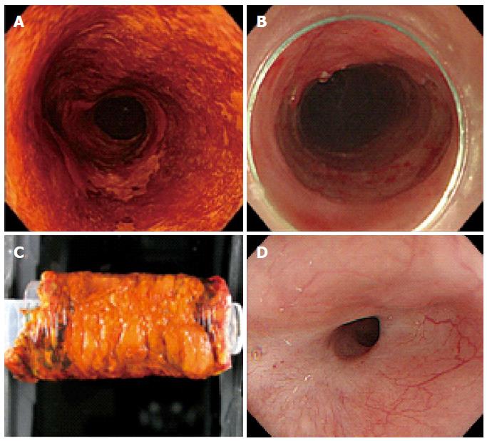
Figure 1 Representative endoscopic imaging of a stricture after circumferential endoscopic submucosal dissection for a large-sized superficial esophageal squamous cell carcinoma.
A: Lugor chromoendoscopy imaging; B: Circumferential post-ESD ulceration; C: ESD specimen; D: Endoscopic imaging of post-ESD stricture 3 mo after ESD. ESD: Endoscopic submucosal dissection.
- Citation: Uno K, Iijima K, Koike T, Shimosegawa T. Useful strategies to prevent severe stricture after endoscopic submucosal dissection for superficial esophageal neoplasm. World J Gastroenterol 2015; 21(23): 7120-7133
- URL: https://www.wjgnet.com/1007-9327/full/v21/i23/7120.htm
- DOI: https://dx.doi.org/10.3748/wjg.v21.i23.7120


