Copyright
©The Author(s) 2015.
World J Gastroenterol. Jun 14, 2015; 21(22): 7052-7058
Published online Jun 14, 2015. doi: 10.3748/wjg.v21.i22.7052
Published online Jun 14, 2015. doi: 10.3748/wjg.v21.i22.7052
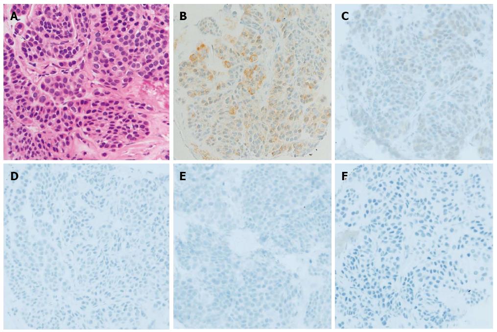
Figure 2 Endoscopic ultrasound-guided fine-needle aspiration biopsy pathology.
A: Proliferating oval-shaped cells in a small nest formation and a high nuclear cytoplasmic ratio were observed (hematoxylin and eosin stain, × 400 magnification). IHC staining was positive for B: muscle actin, and slightly positive for C: synaptophysin, but negative for D: chromogranin, E: c-kit, F: desmin (× 400).
- Citation: Kato S, Kikuchi K, Chinen K, Murakami T, Kunishima F. Diagnostic utility of endoscopic ultrasound-guided fine-needle aspiration biopsy for glomus tumor of the stomach. World J Gastroenterol 2015; 21(22): 7052-7058
- URL: https://www.wjgnet.com/1007-9327/full/v21/i22/7052.htm
- DOI: https://dx.doi.org/10.3748/wjg.v21.i22.7052


