Copyright
©The Author(s) 2015.
World J Gastroenterol. Jun 14, 2015; 21(22): 6974-6981
Published online Jun 14, 2015. doi: 10.3748/wjg.v21.i22.6974
Published online Jun 14, 2015. doi: 10.3748/wjg.v21.i22.6974
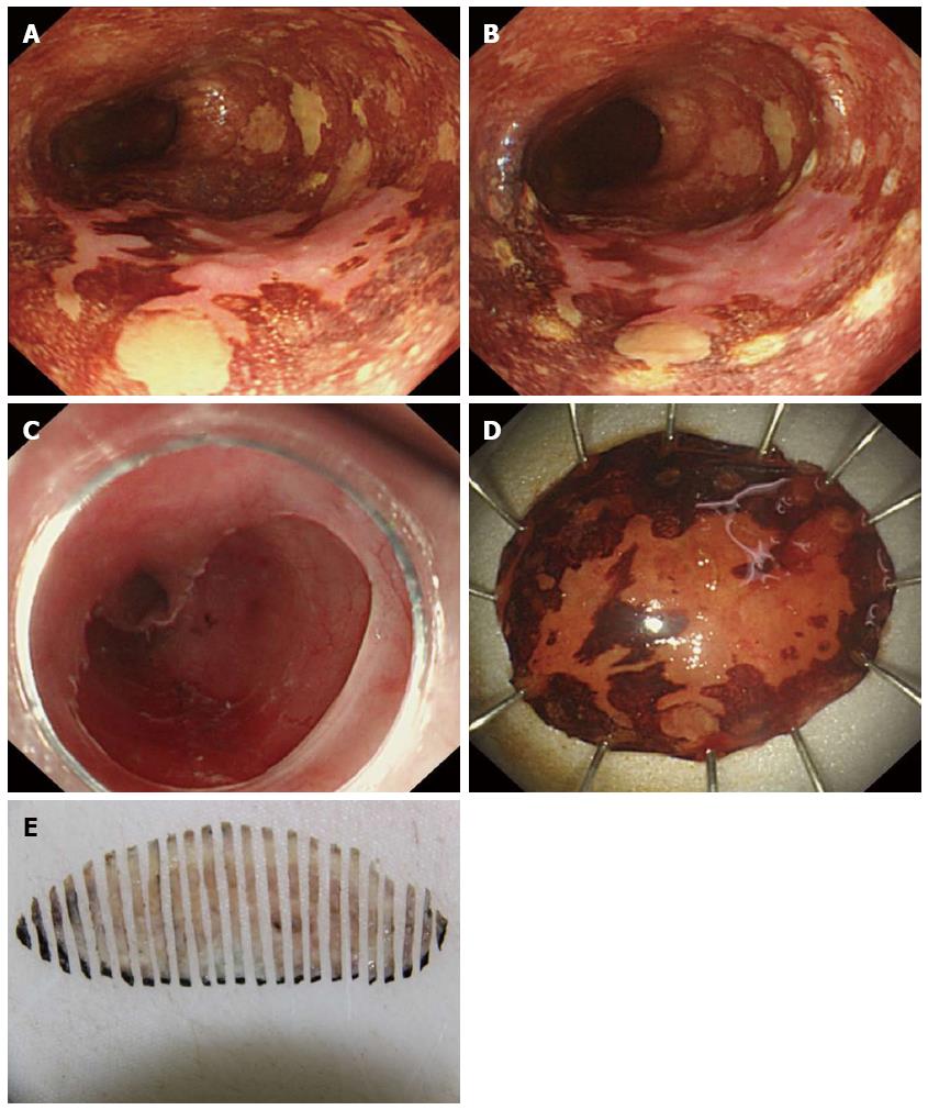
Figure 4 En bloc endoscopic resection for esophageal lesions.
A: Lugol’s iodine chromoendoscopy showed an unstained lesion located in the esophagus; B: The marks surrounding the lesion are at least 5 mm away from the lesion; C: Upper gastrointestinal endoscopy showed an artificial ulceration after ESD; D: En bloc resected specimen. 3.0 cm × 3.0 cm; E: The resected specimen was fixed in 10% formaldehyde solution and continuously sliced into 2 mm sections from the proximal end to the distal end for pathology.
- Citation: Huang J, Yang YS, Lu ZS, Wang SF, Yang J, Yuan J. Detection of superficial esophageal squamous cell neoplasia by chromoendoscopy-guided confocal laser endomicroscopy. World J Gastroenterol 2015; 21(22): 6974-6981
- URL: https://www.wjgnet.com/1007-9327/full/v21/i22/6974.htm
- DOI: https://dx.doi.org/10.3748/wjg.v21.i22.6974


