Copyright
©The Author(s) 2015.
World J Gastroenterol. Jun 14, 2015; 21(22): 6974-6981
Published online Jun 14, 2015. doi: 10.3748/wjg.v21.i22.6974
Published online Jun 14, 2015. doi: 10.3748/wjg.v21.i22.6974
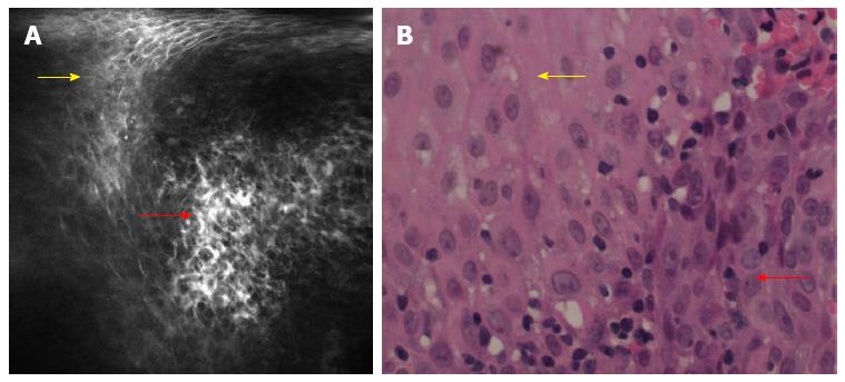
Figure 2 Confocal laser endomicroscopy images of esophageal superficial squamous cell neoplasia.
A: Confocal laser endomicroscopy (CLE) scanning; squamous cells (yellow arrow) are homogeneous, while the indicated squamous cells (red arrow) are irregularly arranged with a distinct size and morphology. Capillary leakage of fluorescein sodium is observed; B: Pathological images; the yellow arrow indicates homogeneous cells; the red arrow indicates disordered cell arrangement. The cells have a distinct size and morphology, which is in accordance with CLE.
- Citation: Huang J, Yang YS, Lu ZS, Wang SF, Yang J, Yuan J. Detection of superficial esophageal squamous cell neoplasia by chromoendoscopy-guided confocal laser endomicroscopy. World J Gastroenterol 2015; 21(22): 6974-6981
- URL: https://www.wjgnet.com/1007-9327/full/v21/i22/6974.htm
- DOI: https://dx.doi.org/10.3748/wjg.v21.i22.6974


