Copyright
©The Author(s) 2015.
World J Gastroenterol. Jun 14, 2015; 21(22): 6872-6883
Published online Jun 14, 2015. doi: 10.3748/wjg.v21.i22.6872
Published online Jun 14, 2015. doi: 10.3748/wjg.v21.i22.6872
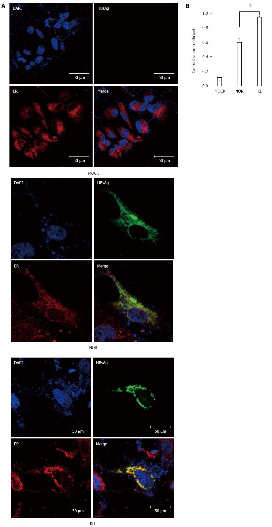
Figure 2 Comparisons of co-localization signals of endoplasmic reticulum and a hepatitis B virus S surface antigen variant (KD) and wild-type hepatitis B virus S surface antigen (NOR) using confocal microscopy.
A: Colocalization of the KD HBsAg variant (KD) and wild-type HBsAg (Nor) in the ER was visualized using confocal microscopy (Confocal A1, Nikon, Japan) after immunofluorescence double-staining assays. HuH-7 cells were transiently transfected with an empty vector (pIRES2-EGFP-Mock) or two different small hepatitis B virus S surface antigen (HBsAg) protein expression vectors (pIRES2-EGFP-Wild-type, pIRES2-EGFP-KD). Cells were harvested 2 d post-transfection, fixed with 4% paraformaldehyde, and stained for HBsAg (green) and the ER marker calnexin (red). The blue color of the nucleus is DAPI staining. Scale bars represent 10, 20, and 50 μm; B: Colocalizations of the HBsAgs and ER in the cytoplasm were compared to each other using co-localization coefficients according to the lasso ROI selection. Statistical comparisons were performed using one-way ANOVA. The average of the coefficients of ten images examined in a double-blinded manner is shown. (bP < 0.01 vs control).
- Citation: Lee IK, Lee SA, Kim H, Won YS, Kim BJ. Induction of endoplasmic reticulum-derived oxidative stress by an occult infection related S surface antigen variant. World J Gastroenterol 2015; 21(22): 6872-6883
- URL: https://www.wjgnet.com/1007-9327/full/v21/i22/6872.htm
- DOI: https://dx.doi.org/10.3748/wjg.v21.i22.6872


