Copyright
©The Author(s) 2015.
World J Gastroenterol. Jun 7, 2015; 21(21): 6759-6763
Published online Jun 7, 2015. doi: 10.3748/wjg.v21.i21.6759
Published online Jun 7, 2015. doi: 10.3748/wjg.v21.i21.6759
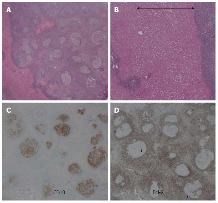
Figure 4 Histological findings.
A: The nodule is well demarcated and comprises a massive infiltration of mature lymphoid cells, forming lymphoid follicles of various sizes with germinal centers (HE staining, low magnification); B: Prominent sinusoidal dilatation (double-headed arrow) seen around the nodule (HE staining, high magnification) is the likely cause of the perinodular enhancement observed on the contrast-enhanced imaging examinations; C, D: Immunohistochemical staining is positive for CD10, and is negative for Bcl-2. These results exclude follicular lymphoma. HE: Hematoxylin and eosin.
- Citation: Sonomura T, Anami S, Takeuchi T, Nakai M, Sahara S, Tanihata H, Sakamoto K, Sato M. Reactive lymphoid hyperplasia of the liver: Perinodular enhancement on contrast-enhanced computed tomography and magnetic resonance imaging. World J Gastroenterol 2015; 21(21): 6759-6763
- URL: https://www.wjgnet.com/1007-9327/full/v21/i21/6759.htm
- DOI: https://dx.doi.org/10.3748/wjg.v21.i21.6759


