Copyright
©The Author(s) 2015.
World J Gastroenterol. Jun 7, 2015; 21(21): 6759-6763
Published online Jun 7, 2015. doi: 10.3748/wjg.v21.i21.6759
Published online Jun 7, 2015. doi: 10.3748/wjg.v21.i21.6759
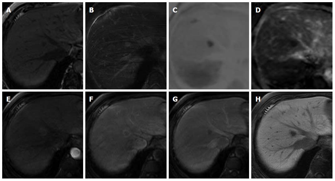
Figure 2 Unenhanced and Gd-EOB-DTPA-enhanced magnetic resonance imaging.
The nodule shows low signal intensity on an unenhanced T1-weighted image (A), and high signal intensity on a T2-weighted image (B). The nodule shows high signal intensity on diffusion-weighted imaging (C) (b = 800 m2/s, inverted black-and-white gray scale), and low signal intensity on apparent diffusion coefficient imaging (D); On Gd-EOB-DTPA-enhanced MRI, the nodule showed perinodular enhancement in the arterial dominant phase (E, F), washout of contrast medium in the late phase (G), and low signal intensity in the hepatobiliary phase (H) suggesting the absence of normal hepatocytes.
- Citation: Sonomura T, Anami S, Takeuchi T, Nakai M, Sahara S, Tanihata H, Sakamoto K, Sato M. Reactive lymphoid hyperplasia of the liver: Perinodular enhancement on contrast-enhanced computed tomography and magnetic resonance imaging. World J Gastroenterol 2015; 21(21): 6759-6763
- URL: https://www.wjgnet.com/1007-9327/full/v21/i21/6759.htm
- DOI: https://dx.doi.org/10.3748/wjg.v21.i21.6759


