Copyright
©The Author(s) 2015.
World J Gastroenterol. Jun 7, 2015; 21(21): 6759-6763
Published online Jun 7, 2015. doi: 10.3748/wjg.v21.i21.6759
Published online Jun 7, 2015. doi: 10.3748/wjg.v21.i21.6759
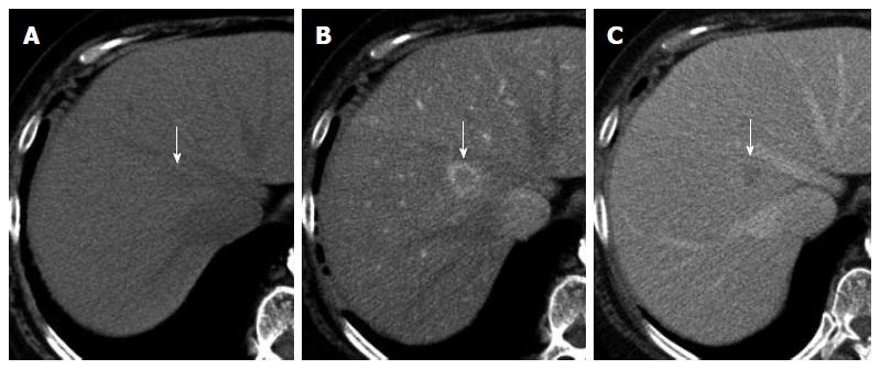
Figure 1 Unenhanced and double-phase contrast-enhanced computed tomography images of the liver, showing a nodule in Segment 1 (arrows).
The nodule shows subtle low attenuation relative to that of liver parenchyma on the unenhanced image (A). On contrast-enhanced computed tomography, the nodule shows perinodular enhancement in the arterial dominant phase (B) and washout of contrast medium in the equilibrium phase (C).
- Citation: Sonomura T, Anami S, Takeuchi T, Nakai M, Sahara S, Tanihata H, Sakamoto K, Sato M. Reactive lymphoid hyperplasia of the liver: Perinodular enhancement on contrast-enhanced computed tomography and magnetic resonance imaging. World J Gastroenterol 2015; 21(21): 6759-6763
- URL: https://www.wjgnet.com/1007-9327/full/v21/i21/6759.htm
- DOI: https://dx.doi.org/10.3748/wjg.v21.i21.6759


