Copyright
©The Author(s) 2015.
World J Gastroenterol. Jun 7, 2015; 21(21): 6582-6590
Published online Jun 7, 2015. doi: 10.3748/wjg.v21.i21.6582
Published online Jun 7, 2015. doi: 10.3748/wjg.v21.i21.6582
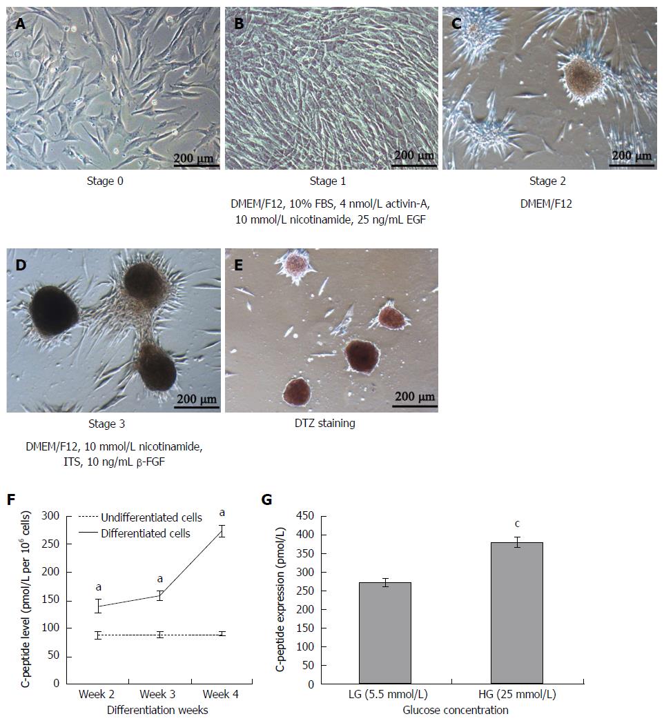
Figure 2 Morphological changes of umbilical cord mesenchymal stem cells during differentiation and C-peptide secretion and stimulation by high glucose levels.
Spindle-shaped and fibroblast-like cells (A, stage 0) were induced to islet-like cluster formation by a 3-stage protocol in 4 wk. After 7 d of induction cells turned round and assembled together (B, stage 1); After another 7 d the round cells became aggregate and some new islet-like clusters began to appear (C, stage 2) and at the end of days 21 mature islet-like clusters with the set of differentiated cells (D, stage 3) that were positive for DTZ staining appeared (E); Spontaneous C-peptide secretion. The media for 106 cells were collected at weeks 2, 3 and 4 and C-peptide concentration were measured (F, aP < 0.05 vs Undifferentiated cells); Glucose challenge test for C-peptide release in response to low (5.5 mmol/L) or high (25 mmol/L) glucose concentration by week 4 differentiated cells (G, cP < 0.05 vs 5.5 mmol/L glucose). Results are the mean ± SE for 6 experiments.
- Citation: Yu YB, Bian JM, Gu DH. Transplantation of insulin-producing cells to treat diabetic rats after 90% pancreatectomy. World J Gastroenterol 2015; 21(21): 6582-6590
- URL: https://www.wjgnet.com/1007-9327/full/v21/i21/6582.htm
- DOI: https://dx.doi.org/10.3748/wjg.v21.i21.6582


