Copyright
©The Author(s) 2015.
World J Gastroenterol. Jun 7, 2015; 21(21): 6582-6590
Published online Jun 7, 2015. doi: 10.3748/wjg.v21.i21.6582
Published online Jun 7, 2015. doi: 10.3748/wjg.v21.i21.6582
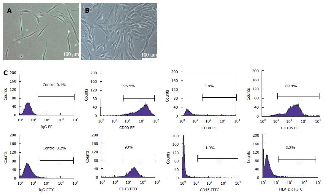
Figure 1 Isolation and identification of umbilical cord mesenchymal stem cells.
Cells displayed spindle-shaped, fibroblast-like morphology (A) and they clonally grew and spread at the bottom of a culture dish within the 12th d (B); Flow cytometric analysis showed that the isolated cells at passage 4 strongly expressed the surface markers of mesenchymal stem cells, such as CD90, CD105 and CD13, but almost no markers of hematopoietic stem cells, such as CD34, CD45 and HLA-DR (C). PE: Phycoerythrin; FITC: Fluorescein isothiocyanate.
- Citation: Yu YB, Bian JM, Gu DH. Transplantation of insulin-producing cells to treat diabetic rats after 90% pancreatectomy. World J Gastroenterol 2015; 21(21): 6582-6590
- URL: https://www.wjgnet.com/1007-9327/full/v21/i21/6582.htm
- DOI: https://dx.doi.org/10.3748/wjg.v21.i21.6582


