Copyright
©The Author(s) 2015.
World J Gastroenterol. Jun 7, 2015; 21(21): 6526-6542
Published online Jun 7, 2015. doi: 10.3748/wjg.v21.i21.6526
Published online Jun 7, 2015. doi: 10.3748/wjg.v21.i21.6526
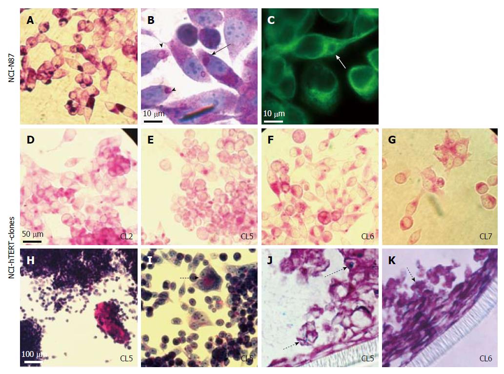
Figure 3 Cell staining analysis for mucins detection on the NCI-N87 parental cell line (A-C) and NCI-hTERT-CL2 (D), CL5 (E, H, I and J), CL6 (F and K) and -CL7 (G).
For neutral mucins detection (stained in pink): PAS-staining (A, D-I); and PAS/haematoxylin staining (B). For acidic mucins detection (J and K) PAS/Alcian blue staining (Alcian positive/PAS negative mucins stained in blue; PAS/Alcian positive mucins stained in purple). α-Tubulin immunodetection (C; green) (1:1000 α-Tubulin mAb plus FITC-conjugated secondary Ab). Black arrow, mucus secreting vesicles that are formed in the endoplasmic reticulum vicinity. Arrow heads, mucus secreting vesicles migrating towards the cytoplasmic membrane and being exocytosed. White arrow, mucus secreting vesicles in close interaction with the microtule network. Dashed arrow suggestive of a more differentiated state for the cells. Dotted arrows, acidic mucins staining.
- Citation: Saraiva-Pava K, Navabi N, Skoog EC, Lindén SK, Oleastro M, Roxo-Rosa M. New NCI-N87-derived human gastric epithelial line after human telomerase catalytic subunit over-expression. World J Gastroenterol 2015; 21(21): 6526-6542
- URL: https://www.wjgnet.com/1007-9327/full/v21/i21/6526.htm
- DOI: https://dx.doi.org/10.3748/wjg.v21.i21.6526


